Services
Services
α-Synuclein Preformed Fibrils (mPFF) Propagation Mouse Model
This model, developed in collaboration with the Michael J. Fox Foundation, enables us to induce propagation of α-synuclein by striatal injection of mouse preformed α-synuclein fibrils. α-synuclein is a key pathological feature of PD and contributes to disease progression.
Synthetic alpha-synuclein pre-formed fibrils (PFF) are capable of ‘seeding’ and propagating alpha synuclein pathology in mice. Intrastriatal PFF inoculation leads to a significant number of alpha-synuclein aggregates that spread over time from the striatum to other brain regions, including dopaminergic neurons of the substantia nigra. PFF inoculation leads to the appearance of Lewy body and Lewy neurite-like inclusions. Over time, the number of fine structured neuritic inclusions reduced with associated neurodegeneration.



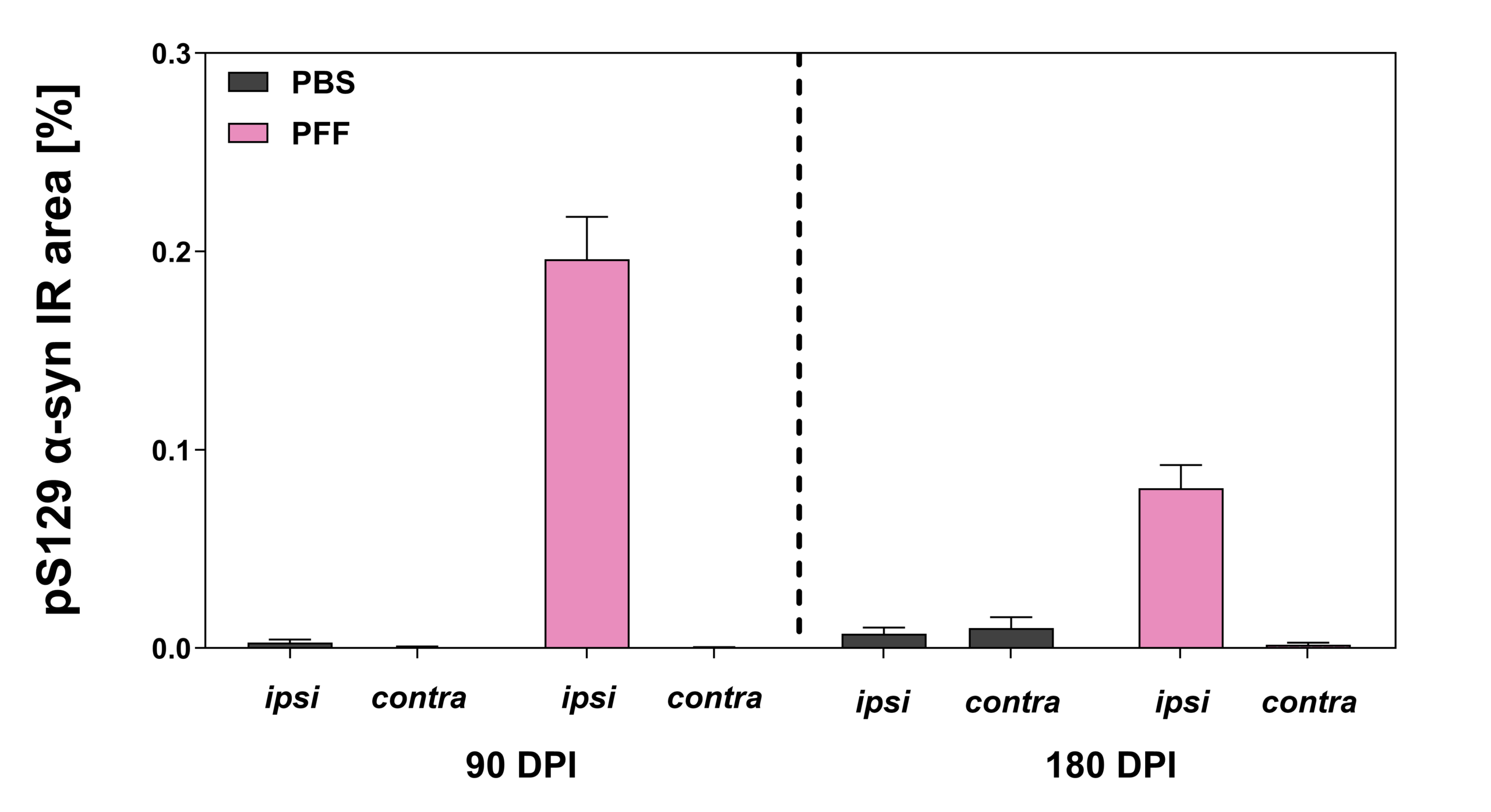
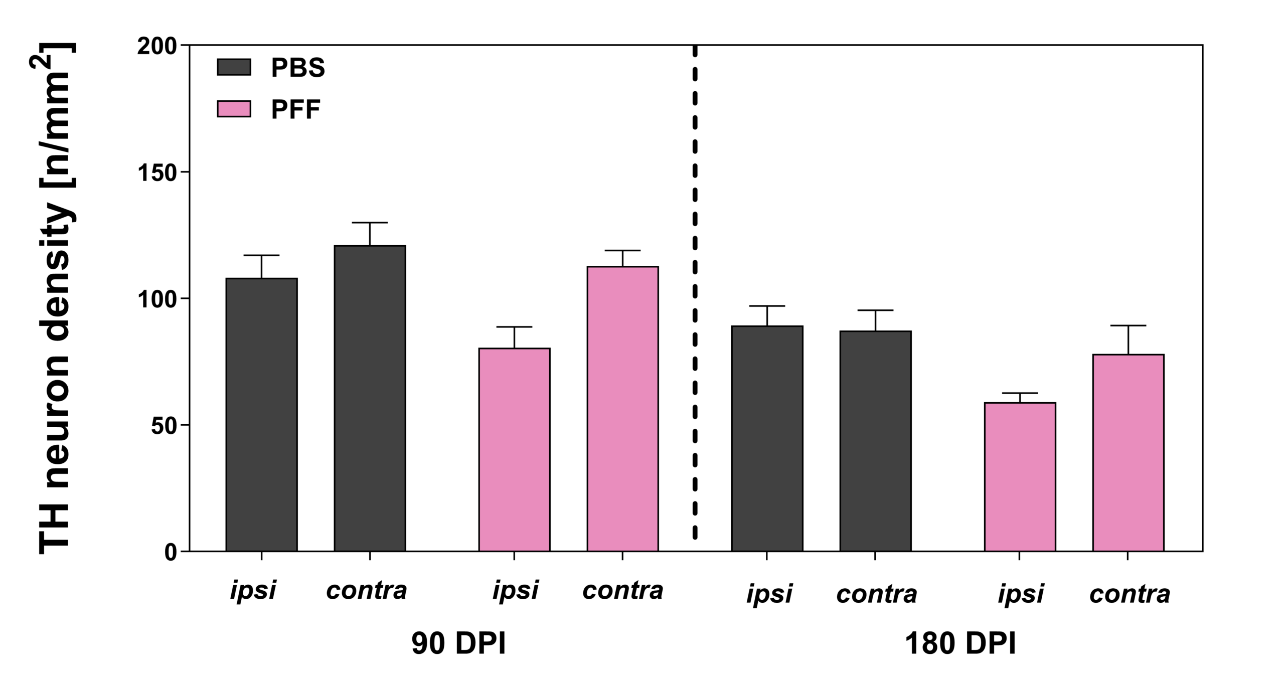
At 90- and 180-days post-PFF injection, a decrease in TH immunoreactivity and cell density was seen in ipsilateral substantia nigra but not contralateral, of PFF-injected mice.
Thy1-α-synuclein (Line 61) Mouse Model







6-OHDA Lesion Rat Model
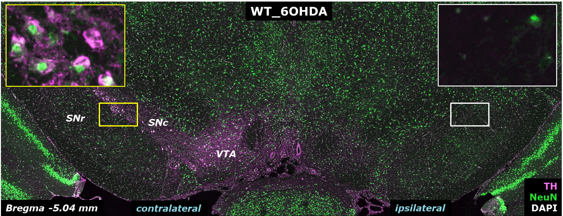
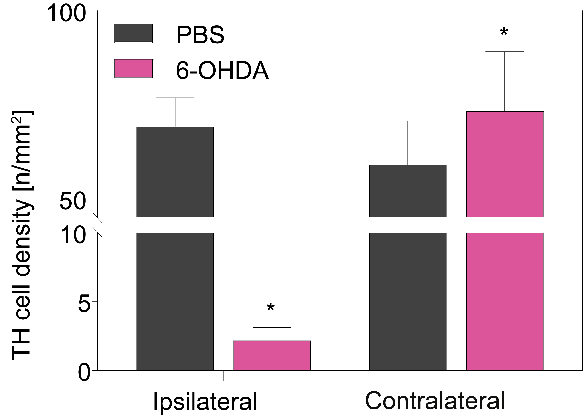
The 6-hydroxy-dopamine (6-OHDA) model simulates PD by targeting and destroying dopaminergic neurons in the brain. Injected 6-OHDA is selectively absorbed by dopamine transporters on neuronal membranes, leading to mitochondrial dysfunction and death. 6-OHDA is delivered unilaterally to the medial forebrain bundle, eliminating dopaminergic connections from the substantia nigra to the striatum. Characteristic outcomes of the model are contralateral motor impairment, decreased striatal dopamine levels, and reduced expression of tyrosine hydroxylase, a critical enzyme in dopamine synthesis.





MPTP Lesion Mouse Model
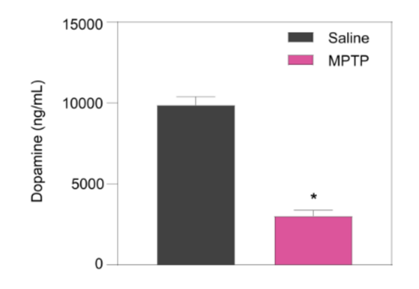
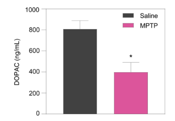
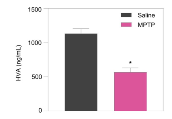
MPTP, or 1-methyl-4-phenyl-1,2,3,6-tetrahydropyridine, is a neurotoxin that selectively damages dopaminergic neurons in the brain. MPTP is administered intraperitoneally and crosses the blood-brain barrier where it is taken up by dopamine transporters (DAT) on the membranes of dopaminergic neurons.