

Services
Services
Pharmacological modulation of circuit-level inhibition
Services
Services
Services
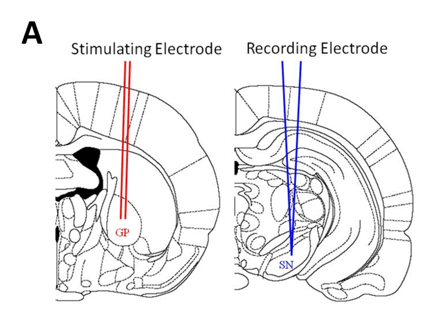
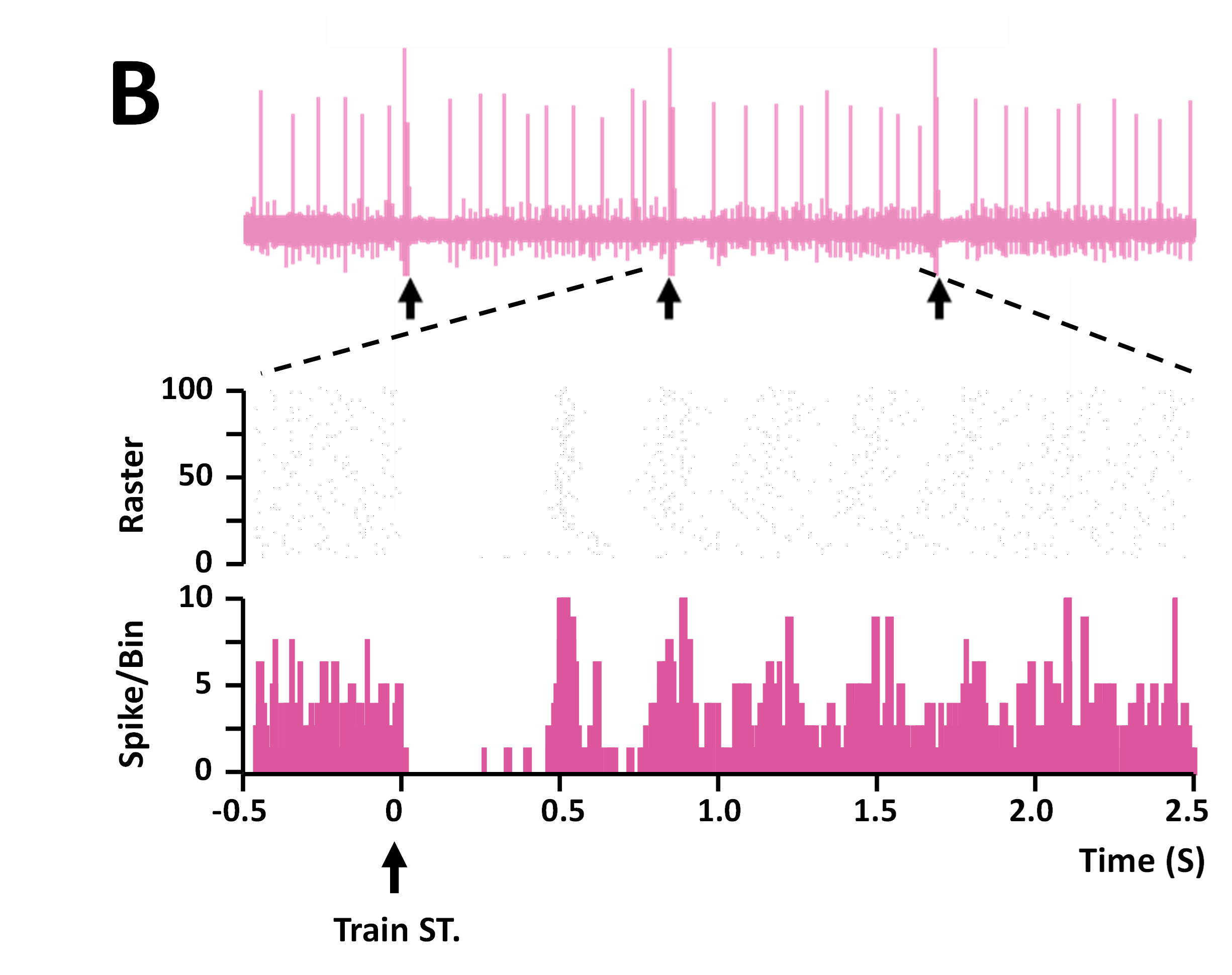
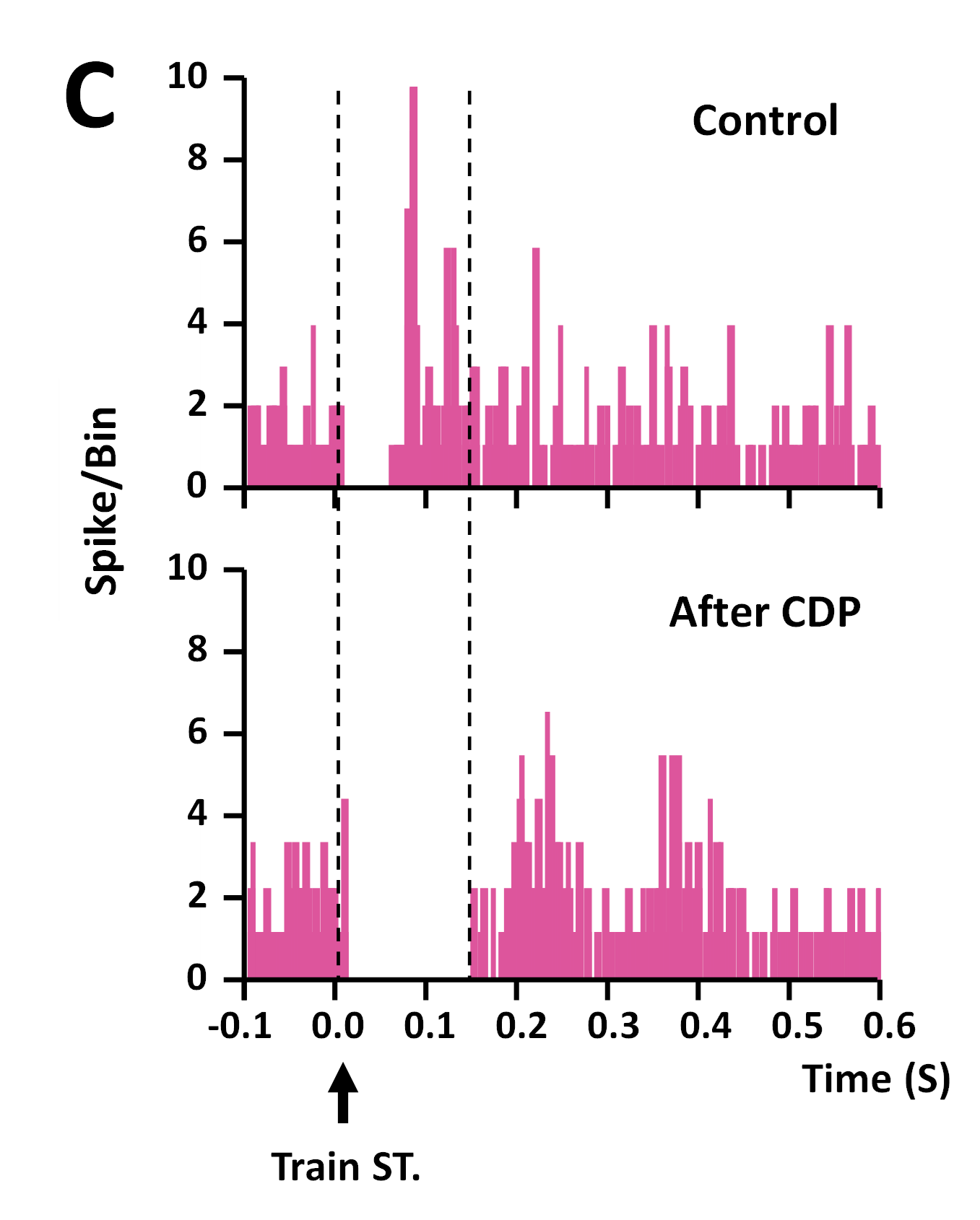
A) Location of stimulating (within the GP) and recording (within the SN) electrodes. B) Top – example of spike trace recorded in SN showing neuronal firing. Each spike represents a single neuronal firing event. Black arrows indicated stimulus trains delivered to the GP, which causes a transient cessation of spike firing within the SN. Middle – raster plot of firing activity from area of top trace as indicated by dashed lines. Bottom – Spike firing binned by time, clearly showing the temporary cessation of activity in the SN, an effect caused by train stimulation of the GP (as indicated by black arrow). C) Chlordiazepoxide (CDP, 5 mg/kg i.v.), a positive allosteric modulator of GABA-A receptors, expands the duration of inhibition of SN firing (bottom trace) produced by GP stimulation compared to control (top trace).
Pharmacological modulation and monitoring of single neuron activity
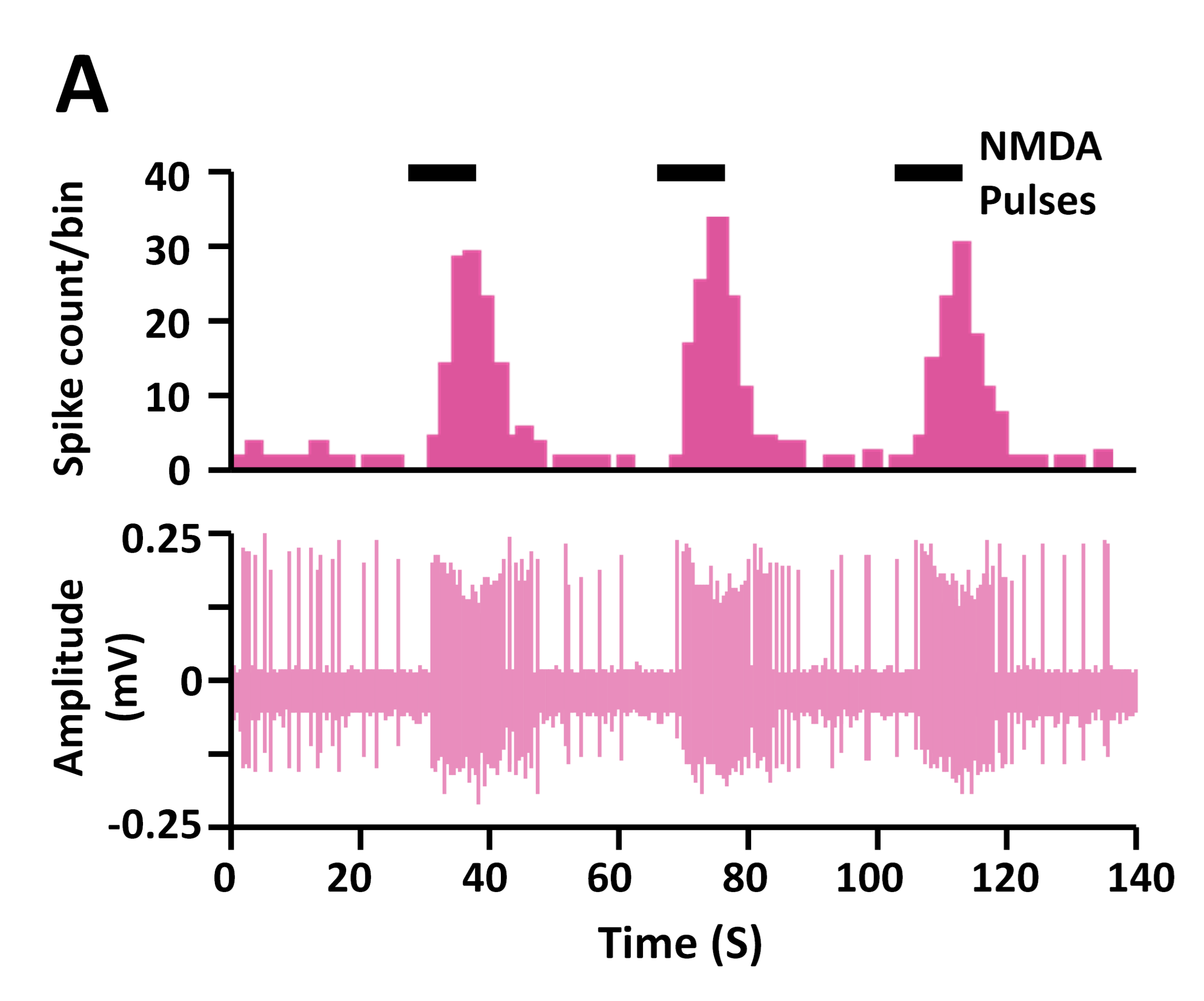
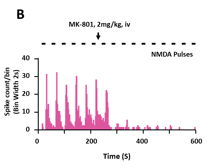
NMDA-induced activation of a single neuron in rat prefrontal cortex (PFC) is blocked by MK-801. Microiontophoresis can rapidly deliver low volume drug administration with a rapid application time while bypassing the blood-brain barrier. This can be combined with in vivo recording to measure modulation of single neuron activity. A) Local iontophoretic administration of NMDA (represented by black bars) produces increases in firing frequency (top) of a single neuron in the PFC of an anesthetized rat. Top – spike frequency (binned), bottom – spike amplitude. B) The use-dependent NMDA receptor antagonist MK-801 (2 mg/kg, i.v.) blocks increased neuronal activity produced by iontophoretic NMDA administration, as can be observed by the suppression of spike firing.
Correlation of drug efficacy with drug exposure
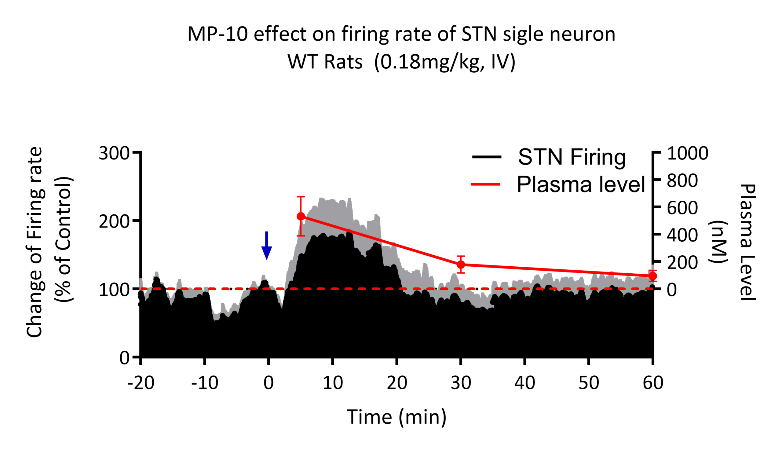
Pharmacodynamic-exposure relationships can be established by monitoring neuronal activity over time and correlating it with drug levels in serially collected plasma samples. Here we show the relationship between plasma concentration of MP-10, a PDE10 inhibitor, and the single unit firing rates of subthalamic nuclei (STN) neurons. The i.v administration of MP-10 produced a marked transient increase in STN firing rate. The rate of firing correlated with the increasing plasma concentration of MP-10; as plasma concentrations of MP-10 decreased over time, so did the mean firing rate of STN neurons. Blue arrow represents the time of MP-10 administration. The red dotted line represents the baseline firing rate of the STN neurons when no compound is present. The grey shading represents the standard error of STN firing.