

Services
Services
LONG-TERM POTENTIATION (LTP)
LTP deficits observed in females of a mouse model of Rett (RTT) syndrome, MeCP2tm1.1 Bird
Services
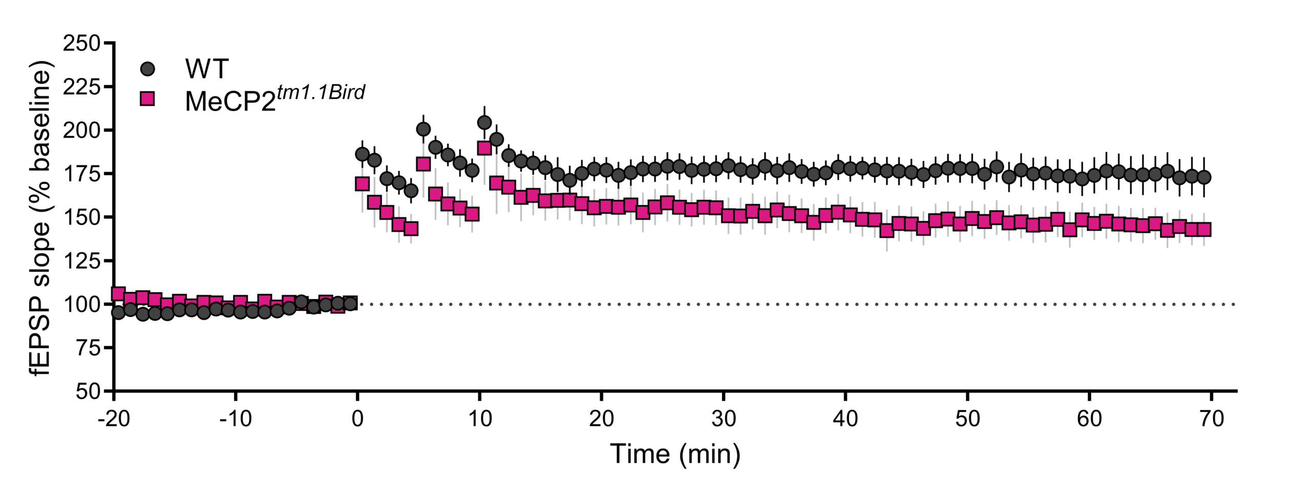
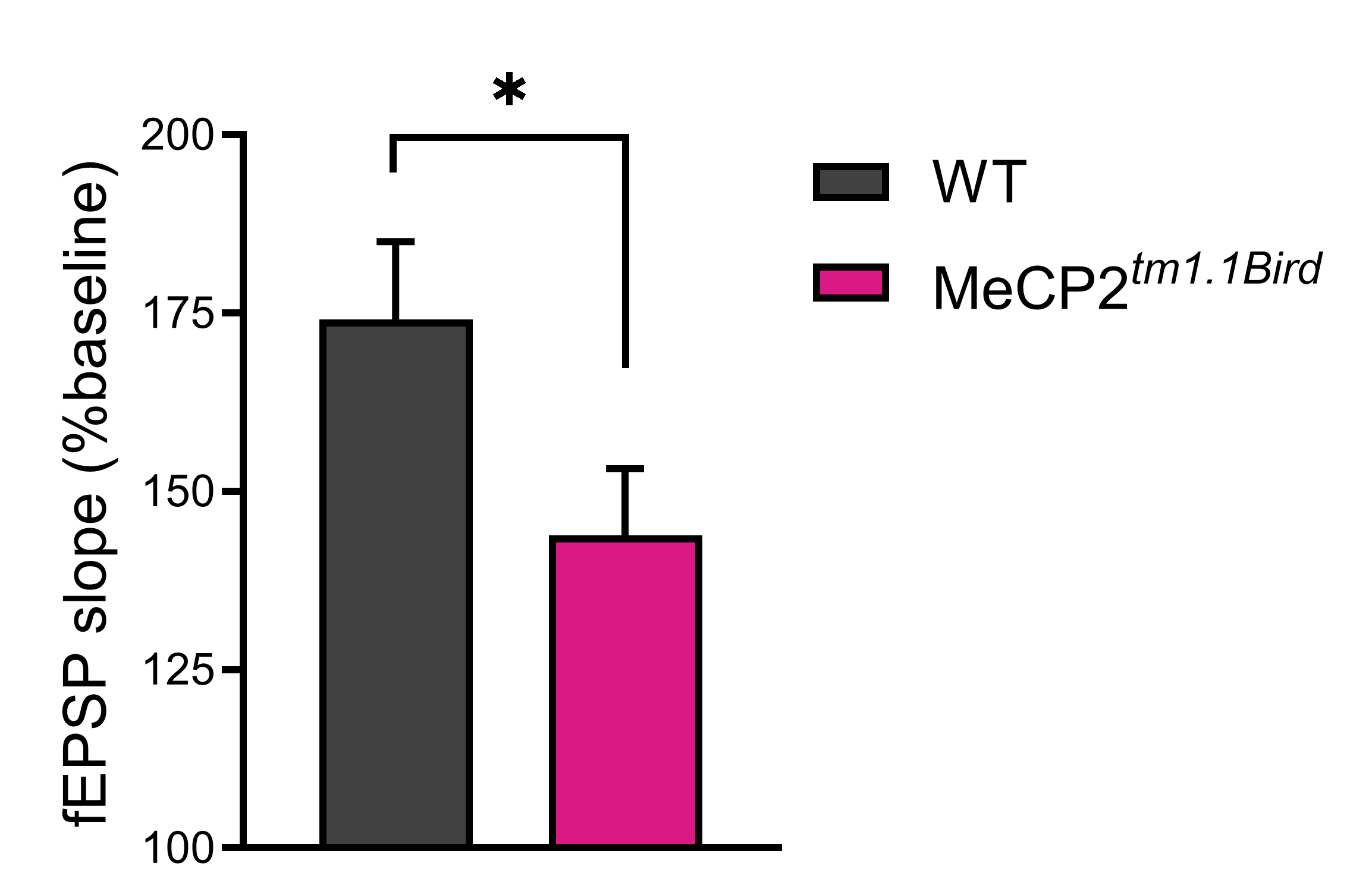
(Top) Extracellular field excitatory postsynaptic potentials (fEPSP) were evoked in hippocampal slices at the Schaffer collateral-CA1 synapse from 6-month-old female wild-type (WT) (n=14 slices from 5 mice) and MeCP2tm1.1Bird (n=11 slices from 5 mice). Stable baseline recordings were obtained with fEPSPs evoked using 40% of maximum stimulus intensity. LTP was induced by delivering three trains of 100 Hz high frequency stimulation, each train separated by five minutes inter-train interval. Synaptic responses were normalized and expressed as percent (%) from baseline. (Bottom) Quantification of the fEPSP slope 65 minutes post-LTP induction showing reduced LTP in MeCP2tm1.1 Bird female mice (p=0.0333, Mann-Whitney Rank Sum Test, two-tailed).
LTP deficits in AD Models:
APP/PS1
Slice electrophysiology: Using extracellular field potential recordings in hippocampal slices, PsychoGenics offers an evaluation of therapeutics designed to improve both pre- and postsynaptic function of APP/PS1 hippocampal CA3-CA1 synapses by evaluating synaptic strength, short- (paired pulse facilitation) and long-term (long term potentiation; LTP) synaptic plasticity.
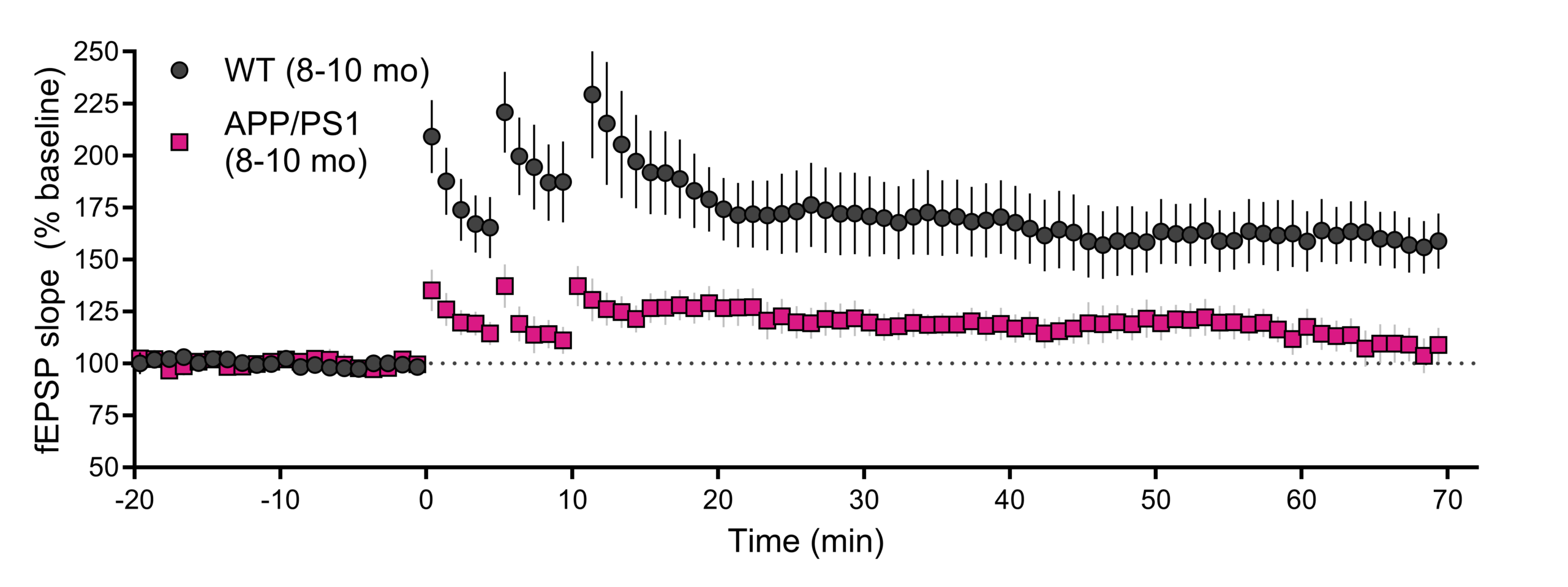
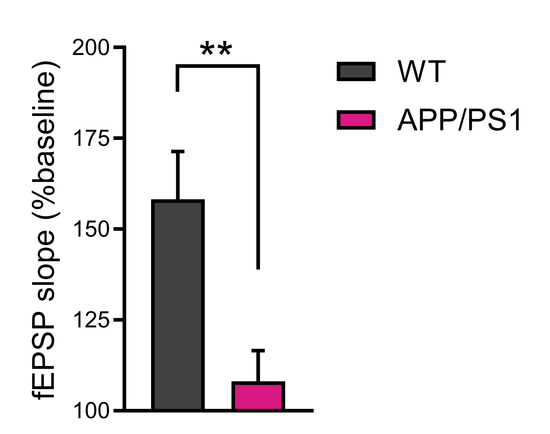
CA3-CA1 long–term potentiation (LTP) is impaired in 8-10 month old APP/PS1 mice. (Top). A time-course of evoked responses (fEPSP slope normalized to the baseline) from an LTP study showing a deficit in APP/PS1 mice. (Bottom). Summary of data taken from the last 5 minutes of recordings (WT: n = 12 slices; APP/PS1: n = 11 slices). Data are presented as mean ± SEM. Data were statistically analyzed with an unpaired t-test (** p < 0.01).
Tg4510
Slice electrophysiology: Our assay offers an evaluation of therapeutics designed to improve both pre- and postsynaptic function of rTg4510 hippocampal CA3-CA1 synapses. Using this approach we can evaluate synaptic strength, short- (paired pulse facilitation) and long-term (e.g. long-term potentiation; LTP) synaptic plasticity. Using multiple approaches we can also differentiate between presynaptic vs postsynaptic changes in synaptic function.
Services
Services
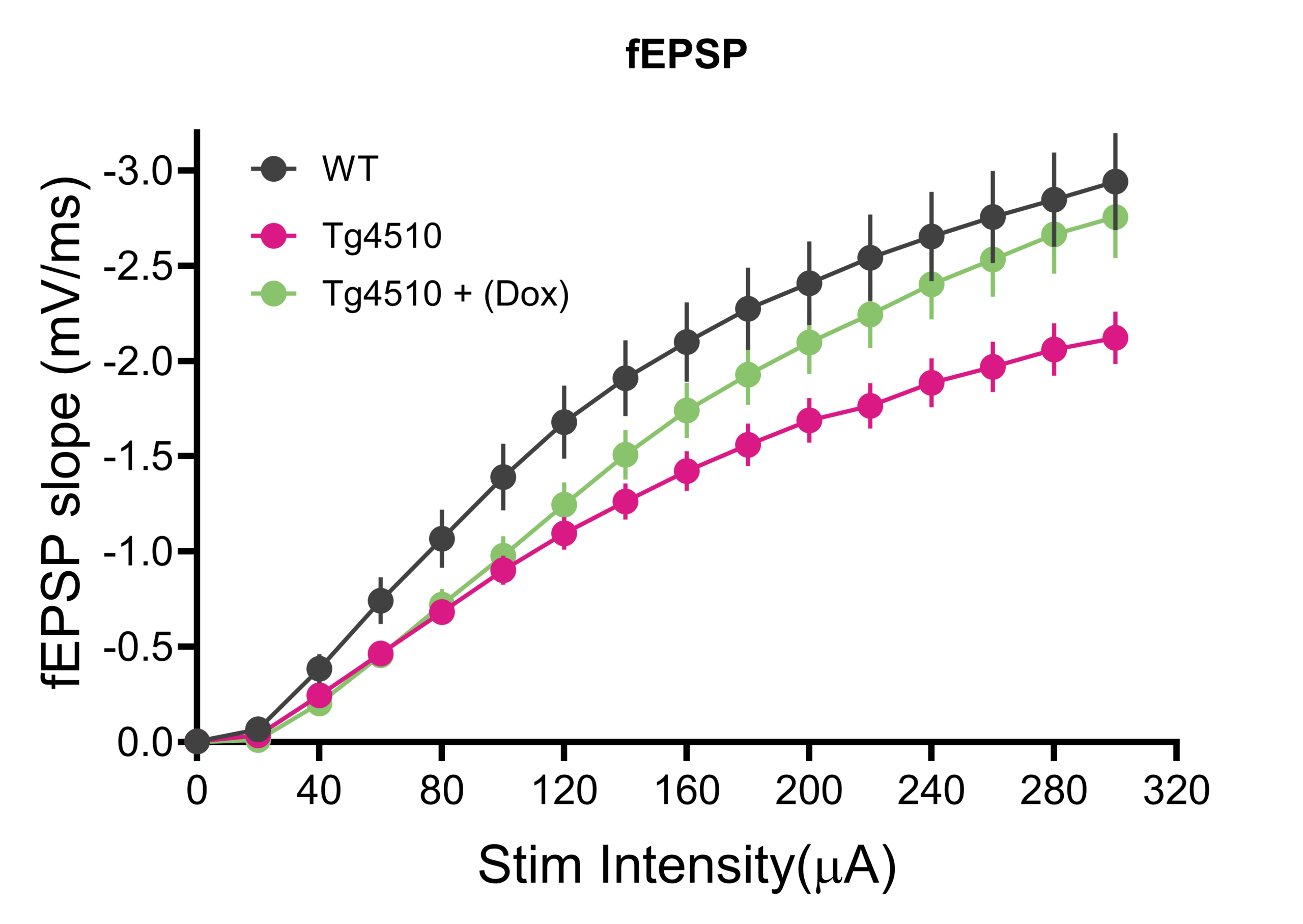
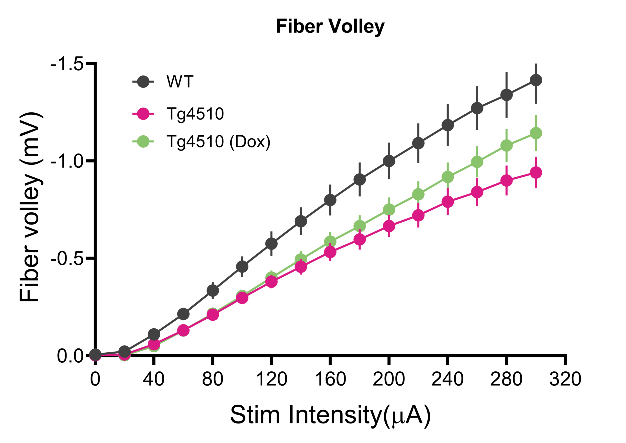
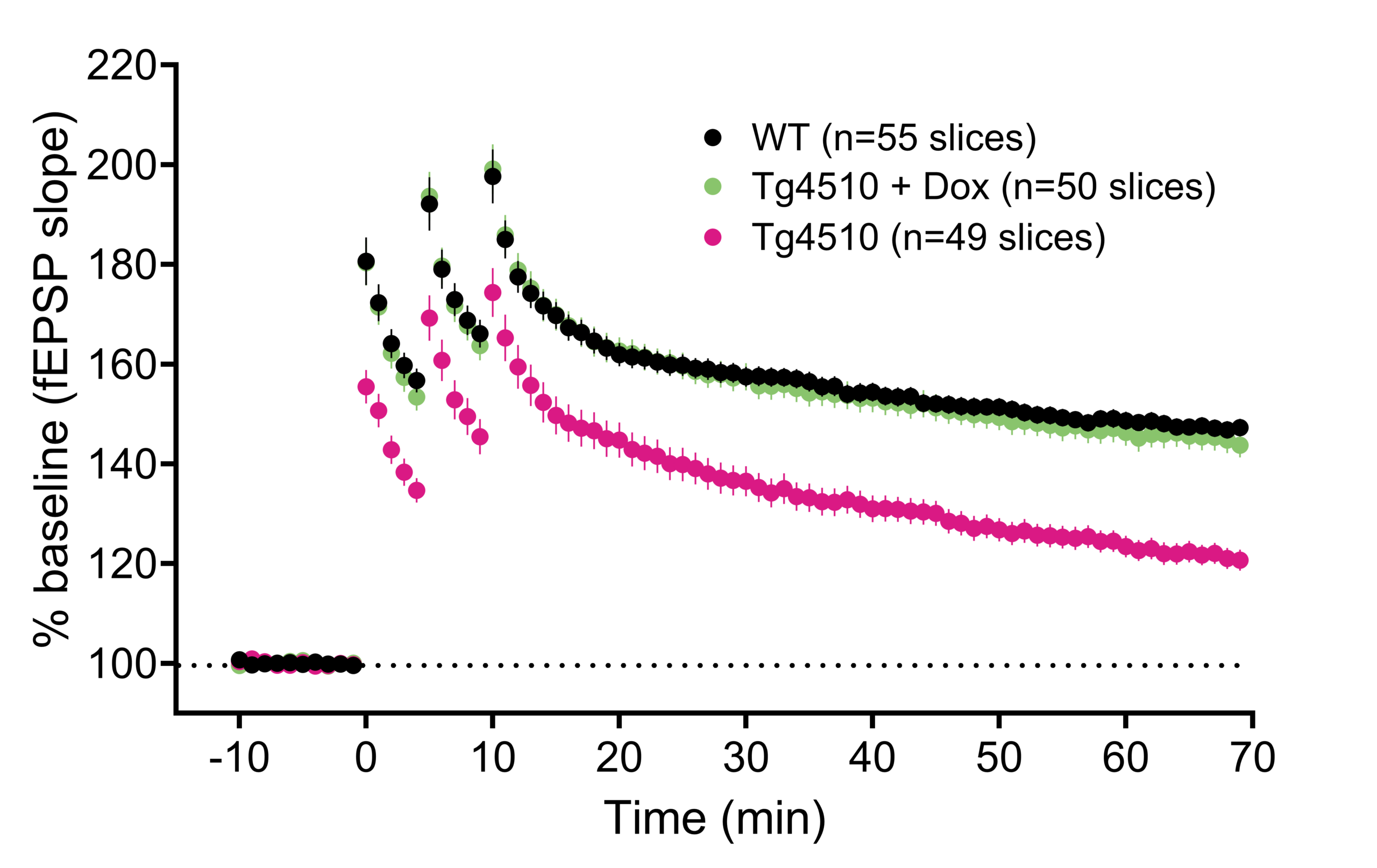
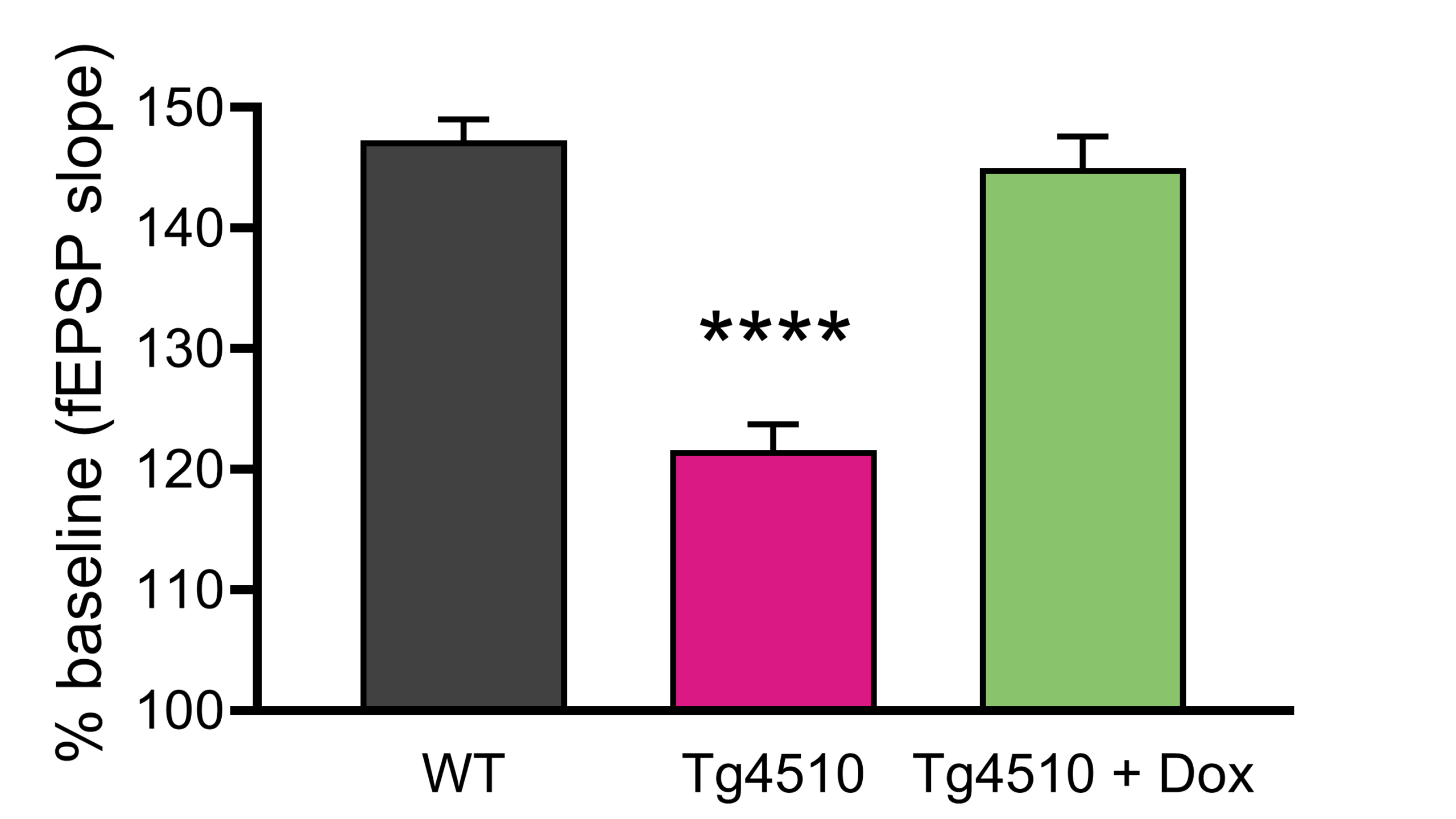
CA3-CA1 long –term potentiation (LTP) is impaired in six-month-old Tg4510 mice. (A) A time-course of evoked responses (fEPSP slope normalized to the baseline) from an LTP study showing a deficit in 6-month-old Tg4510 mice. No LTP deficit was observed in three-month old mice (data not shown), or in six-month-old Tg4510 mice maintained on Dox chow from three months of age. (B) Summary of data taken from the last 5 minutes of recording. Data are presented as mean ± SEM. 1-way ANOVA (****p< 0.0001). A reduction in postsynaptic fEPSP slope (C) and presynaptic fiber volley (D) in Tg4510 hippocampal slices is consistent with overall neurodegeneration. This deficit is partially rescued by Dox.
WHOLE-CELL PATCH-CLAMP ELECTROPHYSIOLOGY
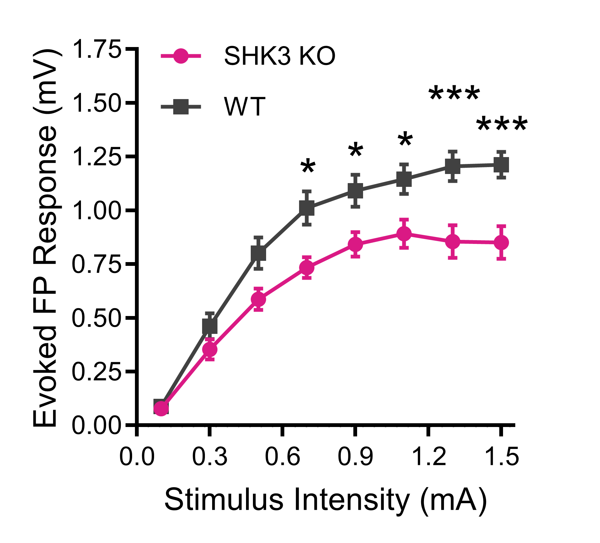
Synaptic transmission deficits in Shank3/F mouse model of ASD:
Mouse
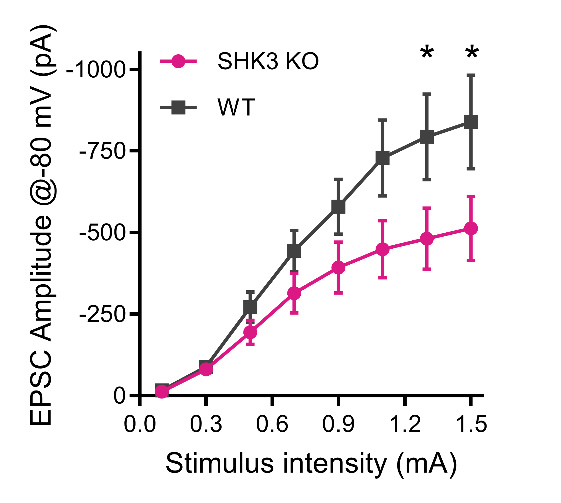
(Top) Synaptically driven extracellular field potentials and (Bottom) whole-cell currents from medium spiny neurons in dorsolateral striatum evoked by stimulating corpus callosum – the major cortical input into basal ganglia – were significantly smaller in Shank3/F KO mice.
Reduced Tonic Inhibition in dentate gyrus observed in male Fragile X mice
Services
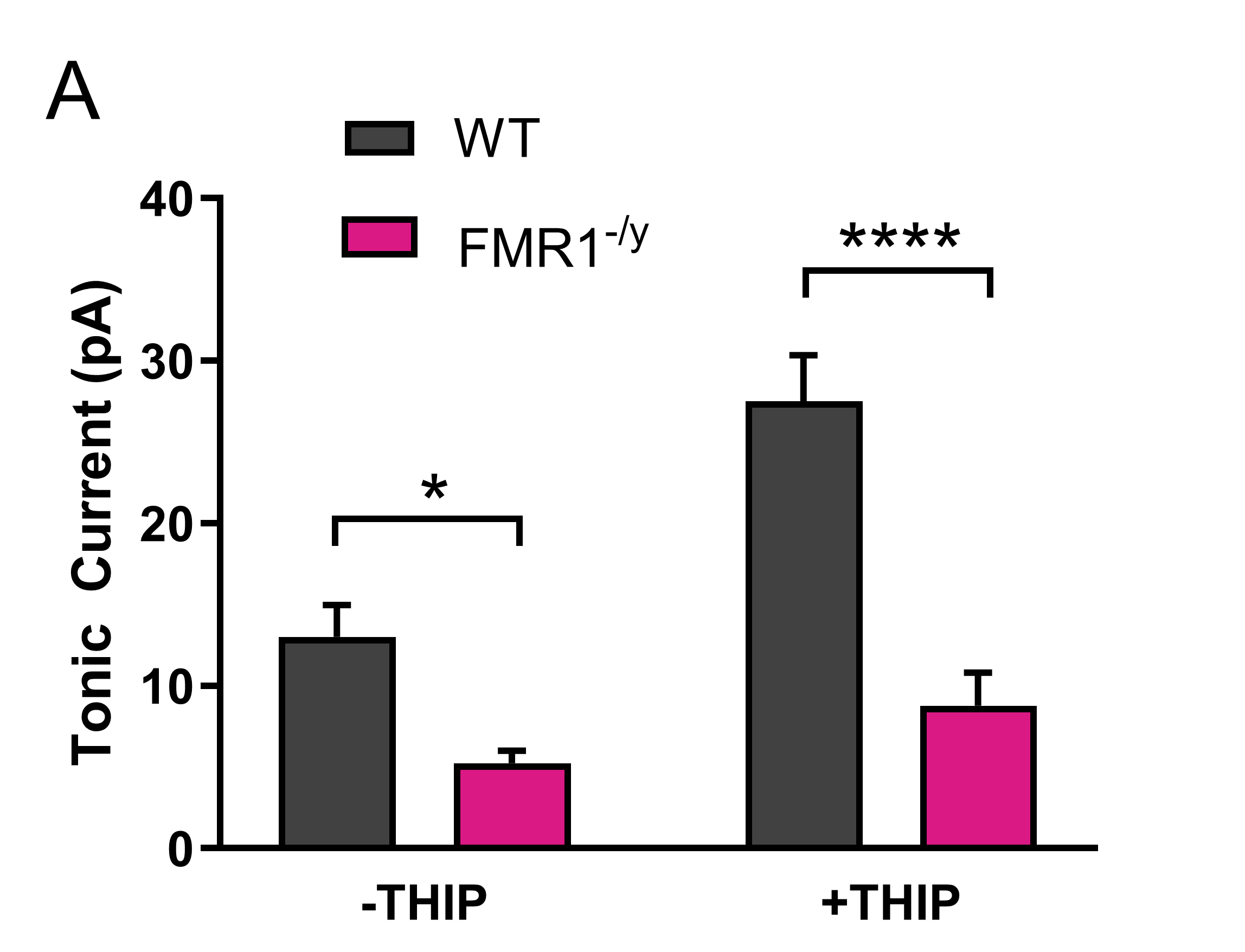
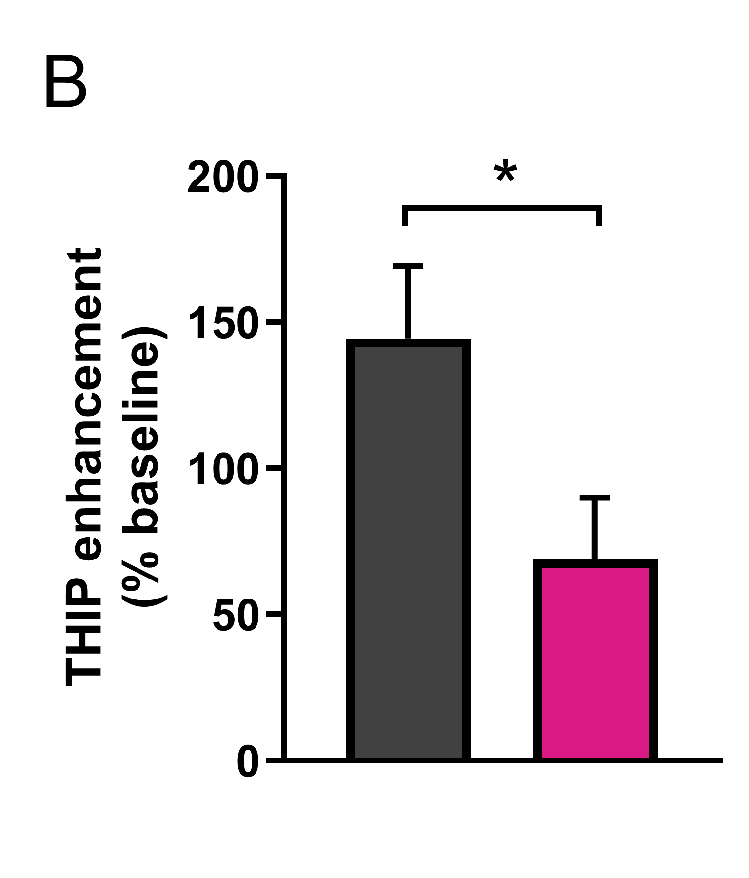
Tonic inhibitory currents are significantly reduced in dentate granule cells of FMR1-/y mice. Whole-cell patch clamp recordings were made in dentate granule cells of hippocampal slices from 2-month-old male wild-type (WT) (n=21 cells, 6 mice) and FMR1-/y mice (n=17 cells, 5 mice), Vhold= -70 mV. (A) Inhibitory tonic current amplitudes at baseline (-THIP) and enhanced by the δ subunit-selective agonist THIP (gaboxadol, 1 μM; +THIP), unmasked by blocking GABAA receptors with 100 μM picrotoxin. *p<0.05, ****p<0.0001, two-way repeated measures ANOVA, Bonferroni’s multiple comparisons test. (B) The degree to which tonic currents can be enhanced by application of THIP is significantly reduced in cells from FMR1-/y mice. n=21 cells (WT), 17 (FMR1-/y), *p<0.05, Mann-Whitney test.
1Kooy, R. et al (2000) Mol. Med. Today; 2Brickley, S. & Mody, I., (2012) Neuron; 3Zhang, N. et al (2017) Exp. Neurol.
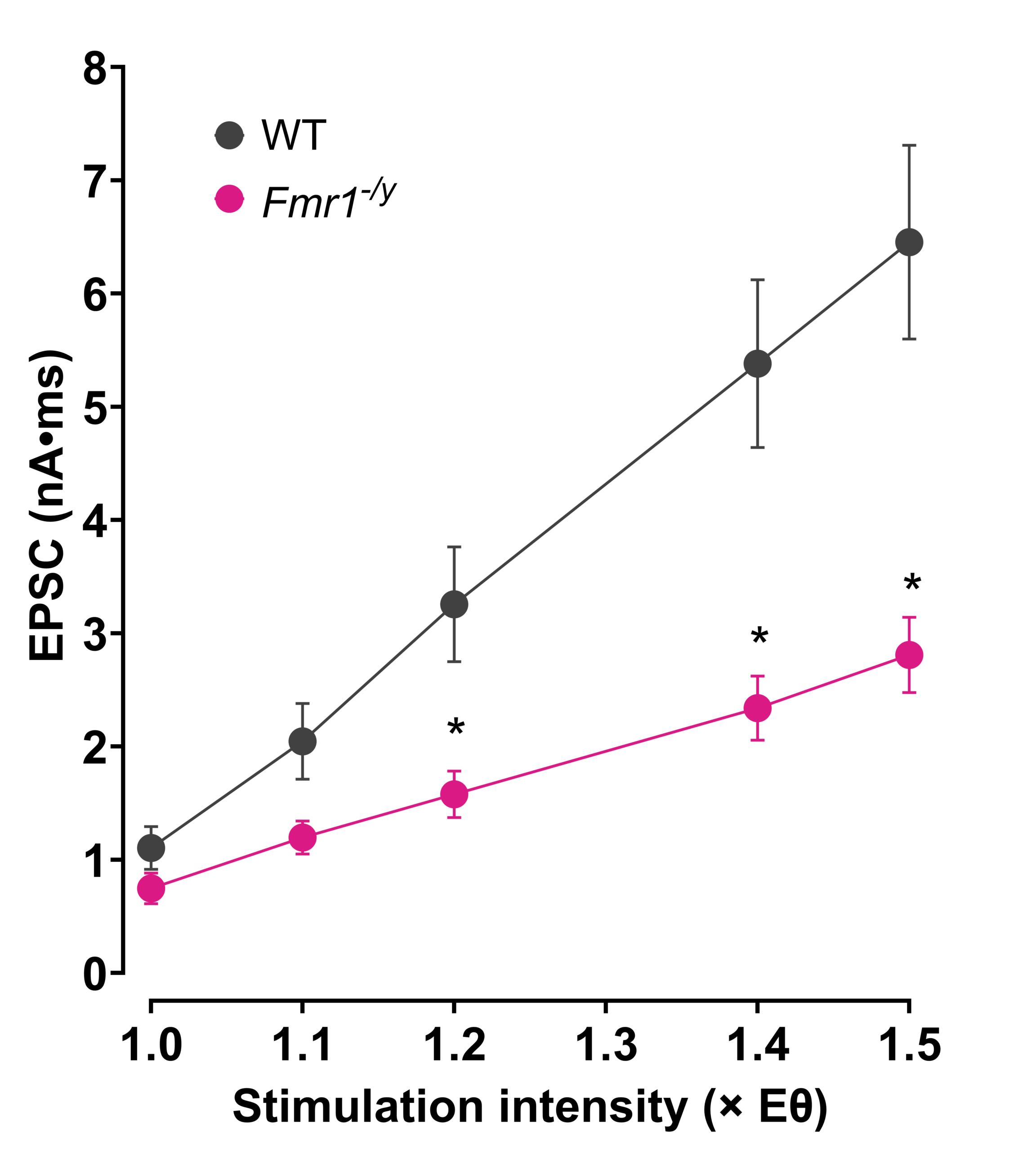
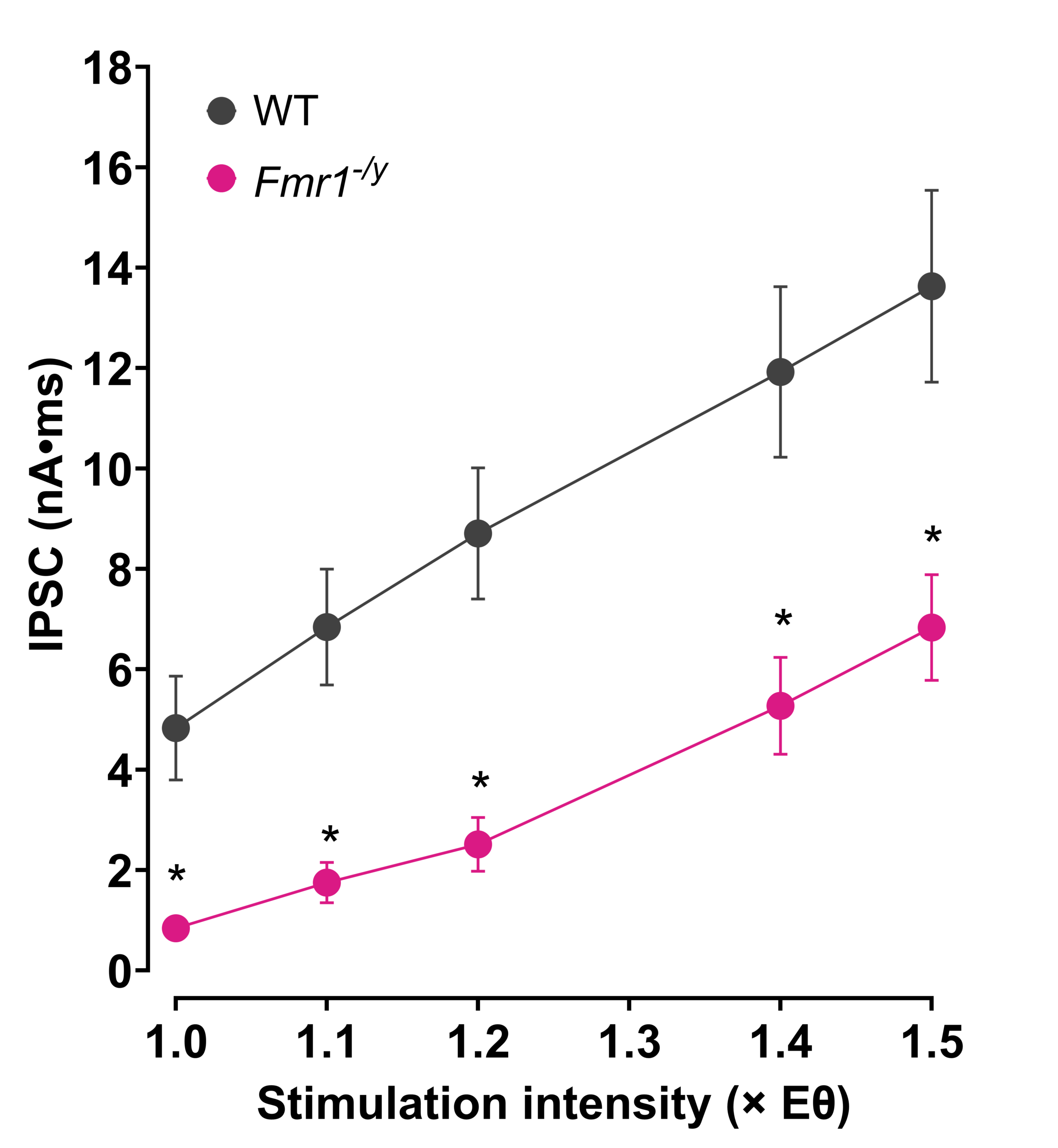
Excitation/Inhibition Imbalances in Fragile X mice: E/I Ratio Shift Favors Excitation in Sensory Cortex
Services
Services
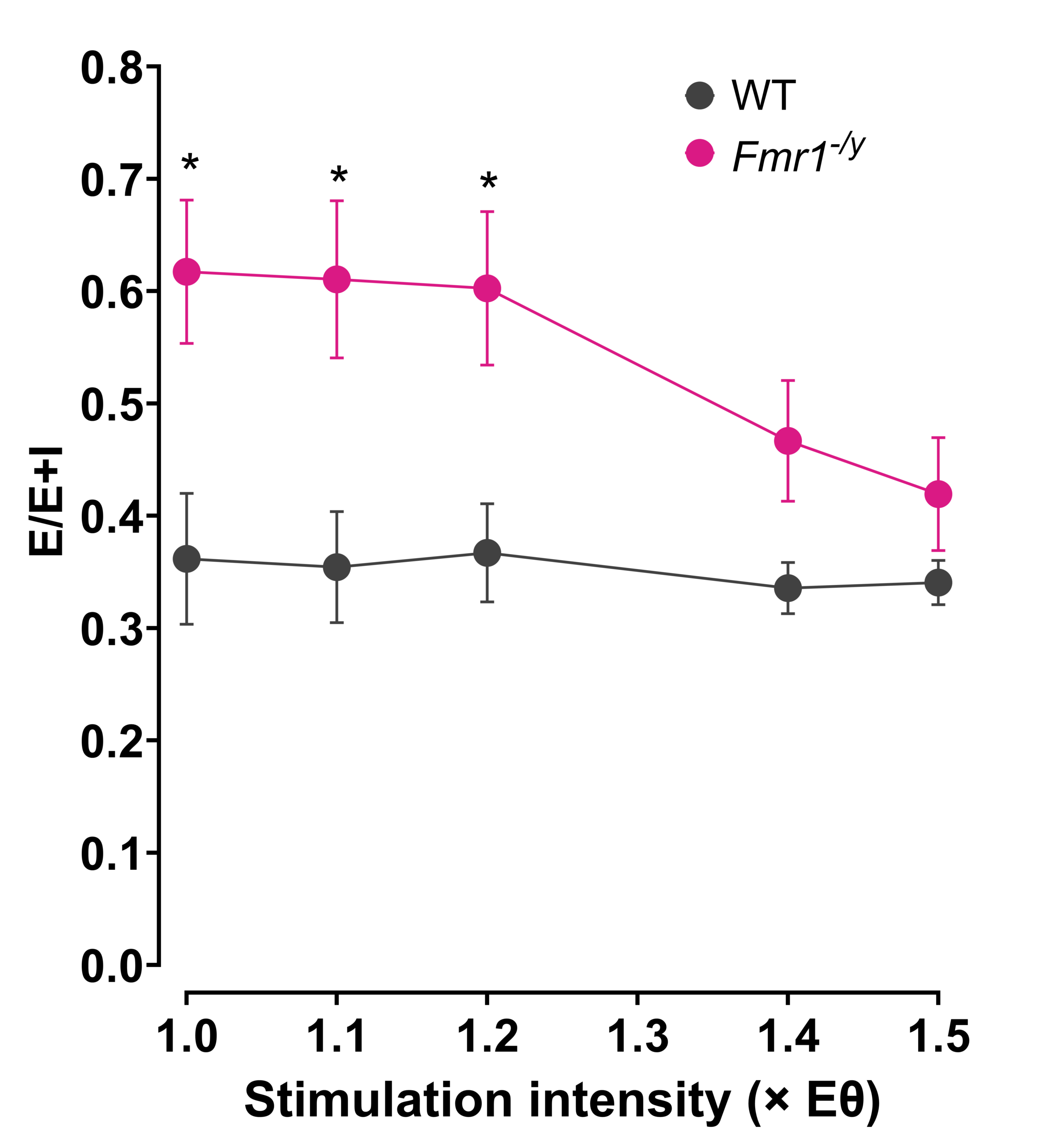
The balance of excitatory and inhibitory neurotransmission shifts in layer II/III pyramidal neurons of sensory cortex. (A) Evoked EPSCs and (B) IPSCs recorded in layer II/III pyramidal cell neurons from WT and FMR1-/y male mice. (C) The resultant ratio of excitatory and inhibitory tone shifts to favor excitation.
Layer 5 motor cortex pyramidal cell hyperexcitability in the TDP-43 (ΔNLS8) mouse model of ALS
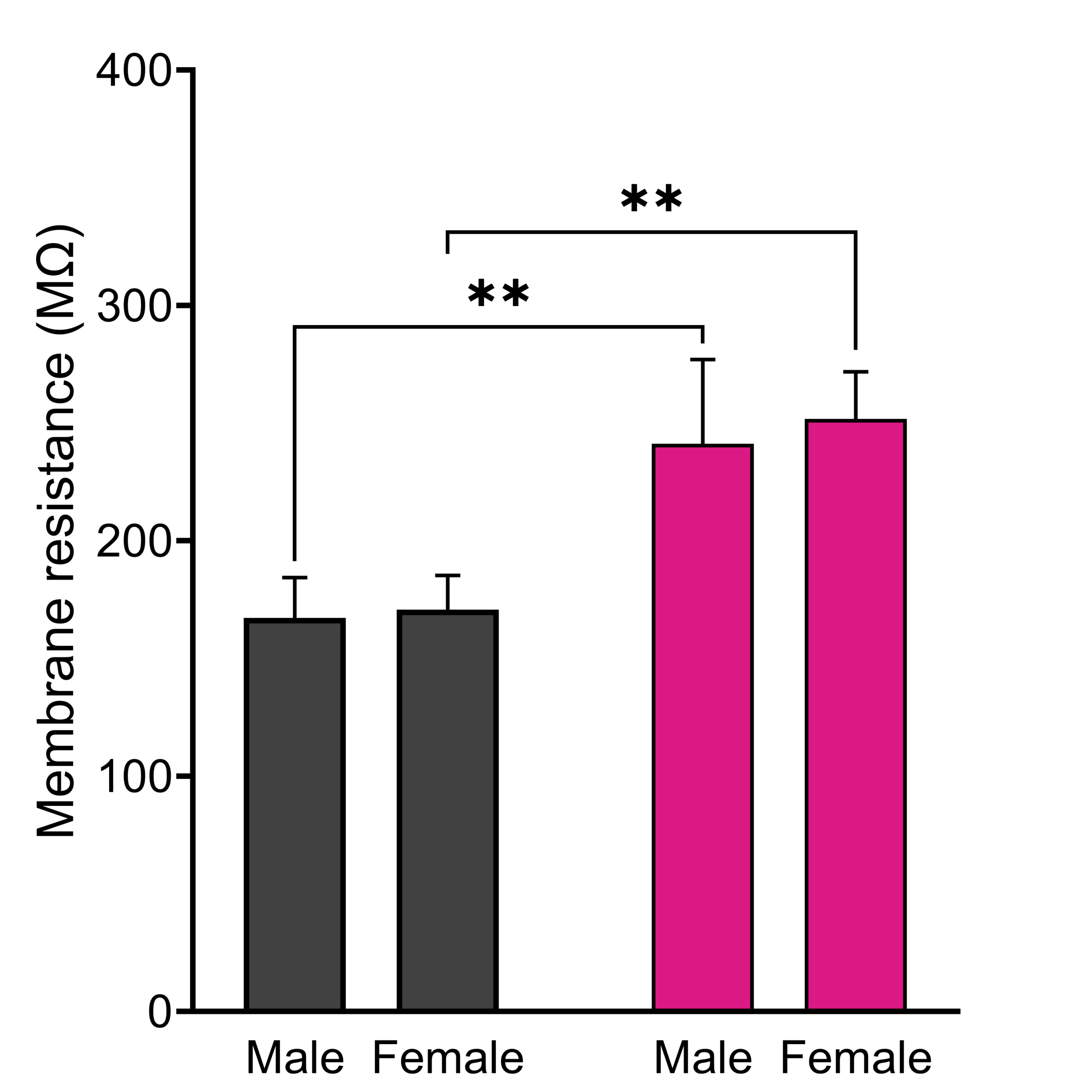
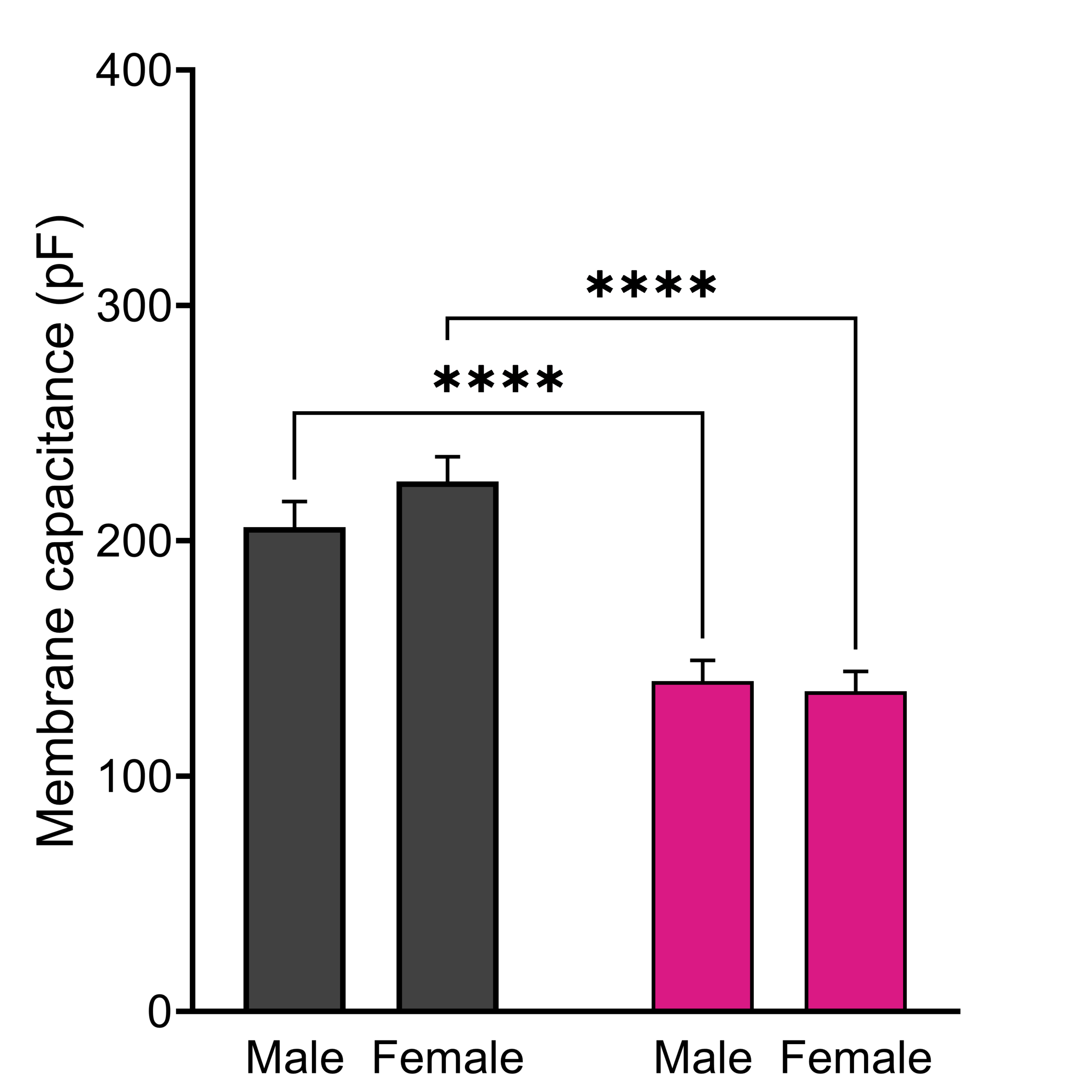
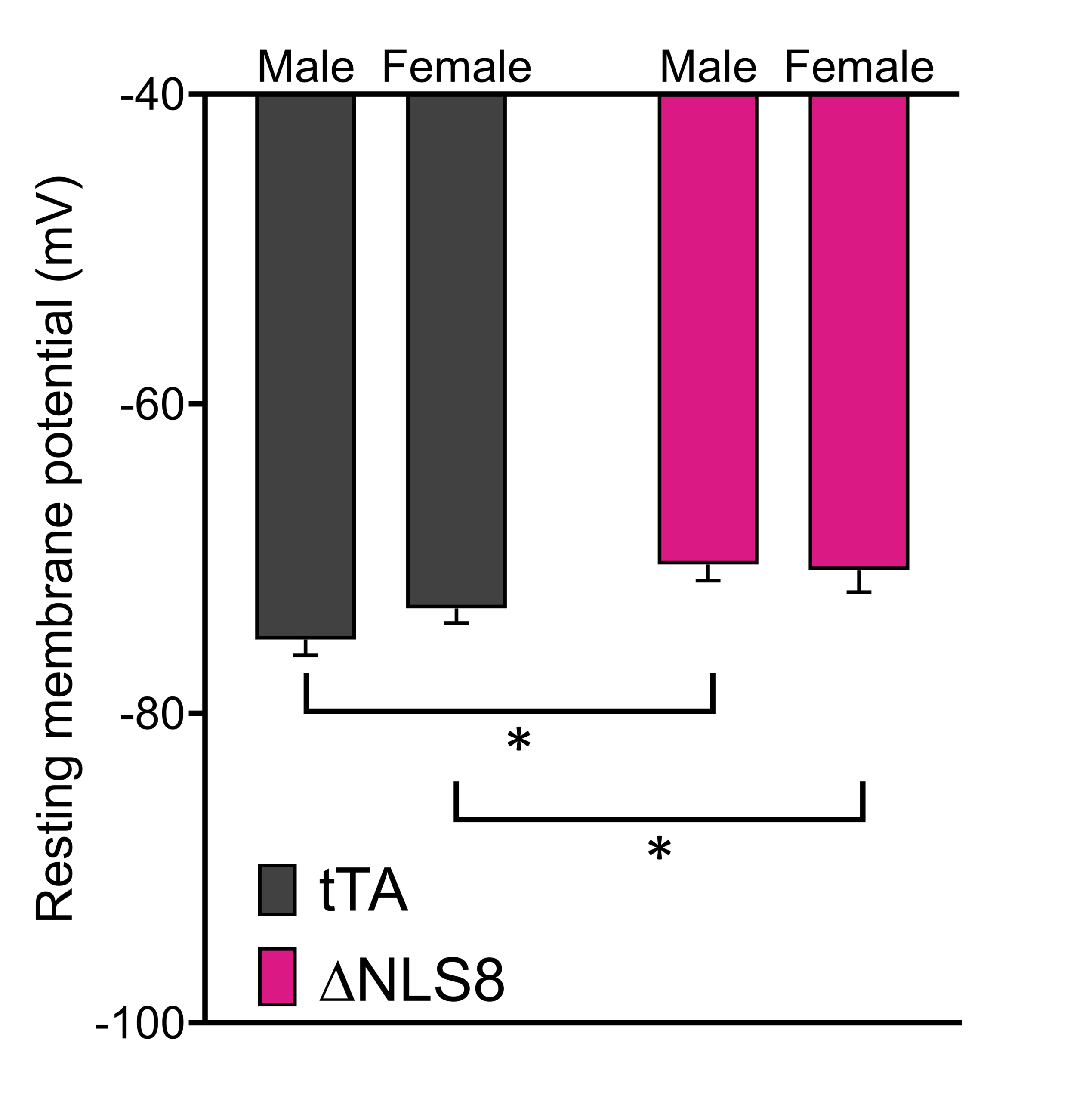
Amyotrophic lateral sclerosis (ALS) is a fatal neurodegenerative disease characterized by progressive loss of upper and lower motor neurons producing muscle denervation, motor impairments and brain atrophy. Deposition of insoluble cytosolic inclusions of TAR-DNA binding protein (TDP-43) correlates with ALS-related pathology in affected tissues. Recent findings have suggested that hyperexcitability of pyramidal cells in the motor cortex, a pathological change observed in both human patients and mouse models of ALS, also occurs in this mouse model1. Psychogenics has confirmed several aspects of this hyperexcitability phenotype in TDP-43 ΔNLS8 mice2, amenable to therapeutic targeting.
Services
Services
1Dyer MS et al, (2021) J Neurochem.; 2Walker AK et al, (2015) Acta Neuropathol.
Services
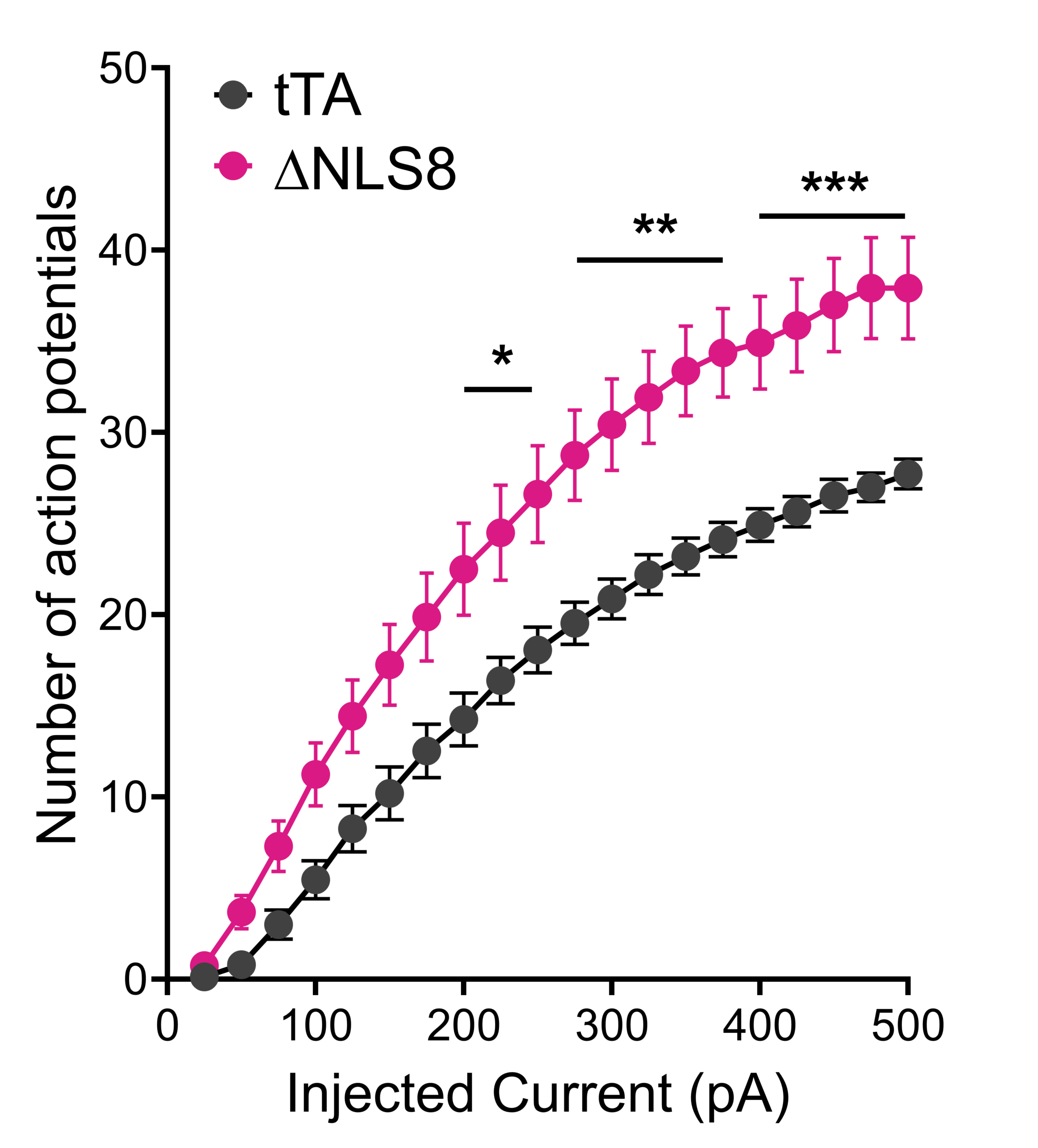
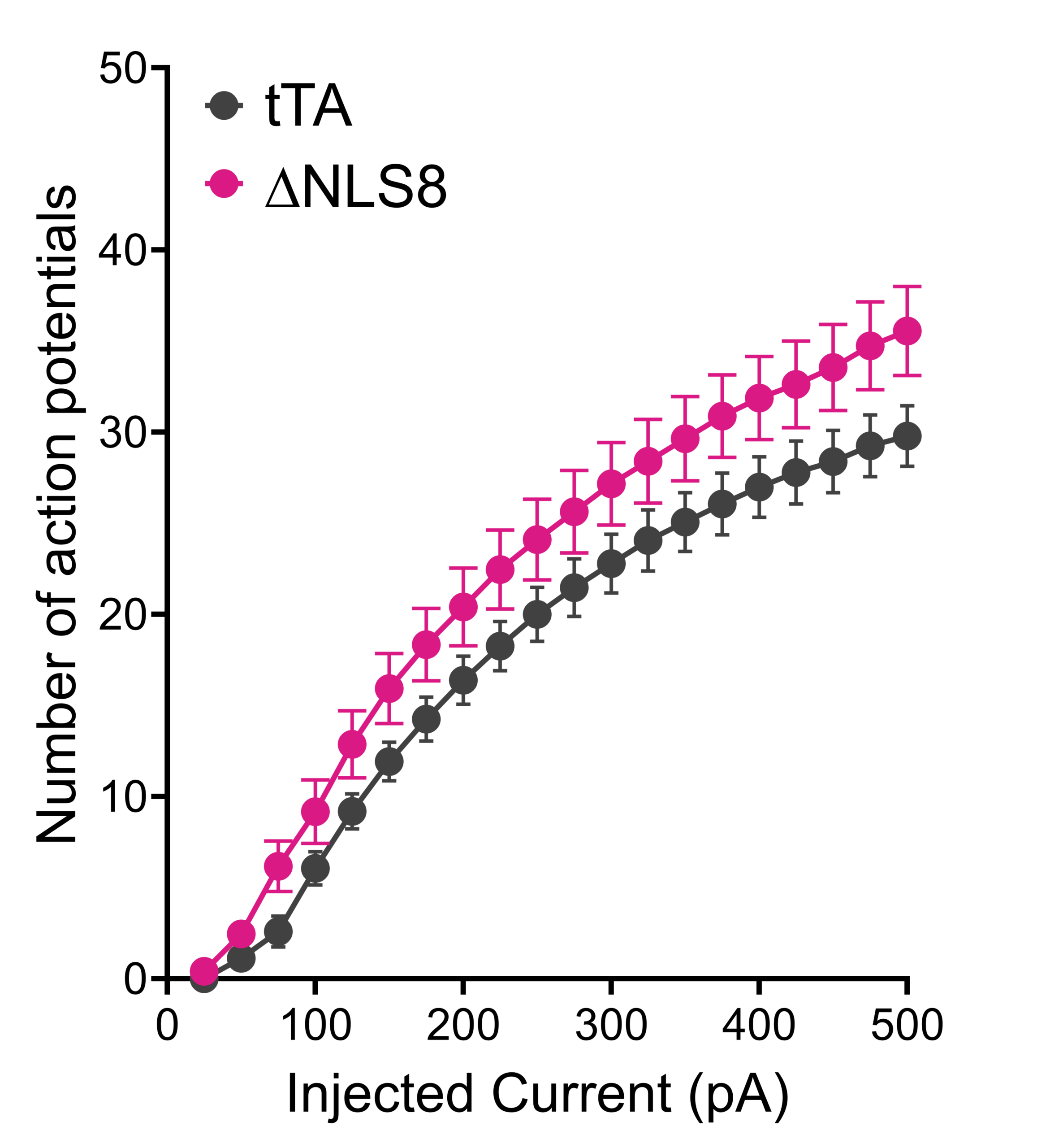
Summary of firing frequency input-output curves. (A) Cells from male ΔNLS8 mice produced a greater number of output action potentials in response to input current injected. Bonferroni’s multiple comparisons test revealed statistically significantly higher action potential numbers at current injection levels of 200 pA and higher; * p<0.05; ** p<0.01; *** p<0.001. (B) Cells from female ΔNLS8 mice showed a trend (p=0.0898) toward greater number of output action potentials in response to input current injected.
Services
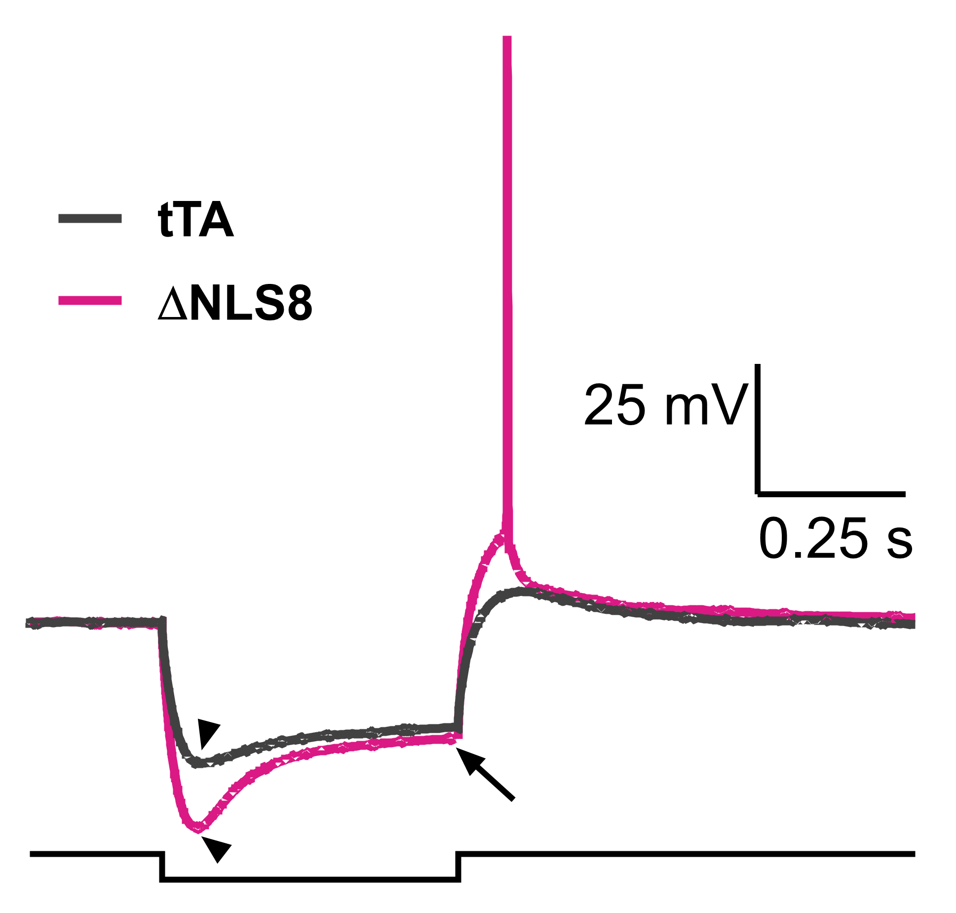
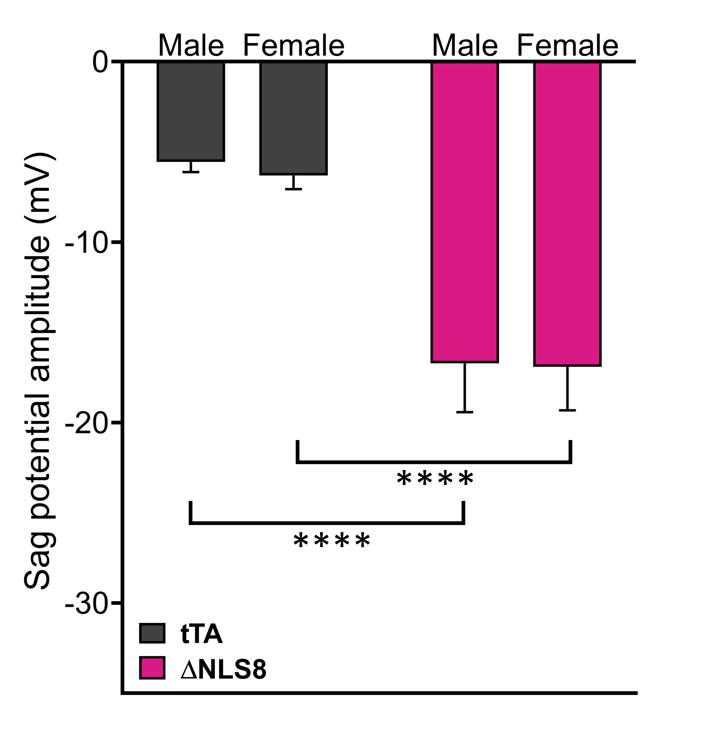
Sag potentials. (A) Example membrane voltage traces (upper traces) of response to injection of -300 pA current pulse (0.5 s; lower square pulse). Cells from ΔNLS8 mice showed slowly developing rebound depolarization – sag potentials of a greater amplitude compared to controls. Sag potential was measured as a difference between the peak potential (arrowheads) and steady-state potential (arrow). Note that in some cases, cells fired “rebound” action potentials upon return from hyperpolarization. (B) Sag potential amplitudes were greater in ΔNLS8 mice of both sexes relative to tTA controls. **** p<0.0001.
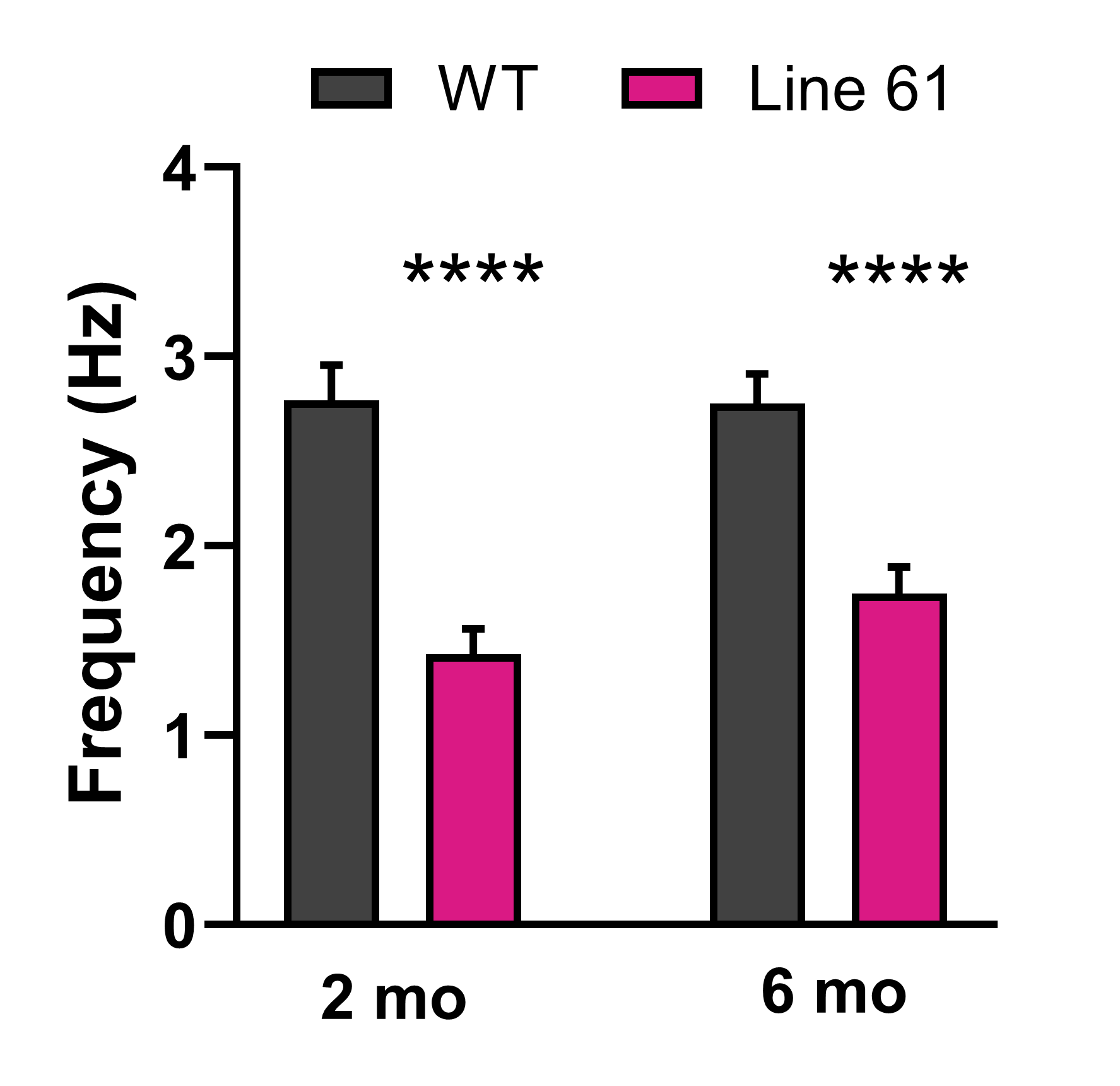
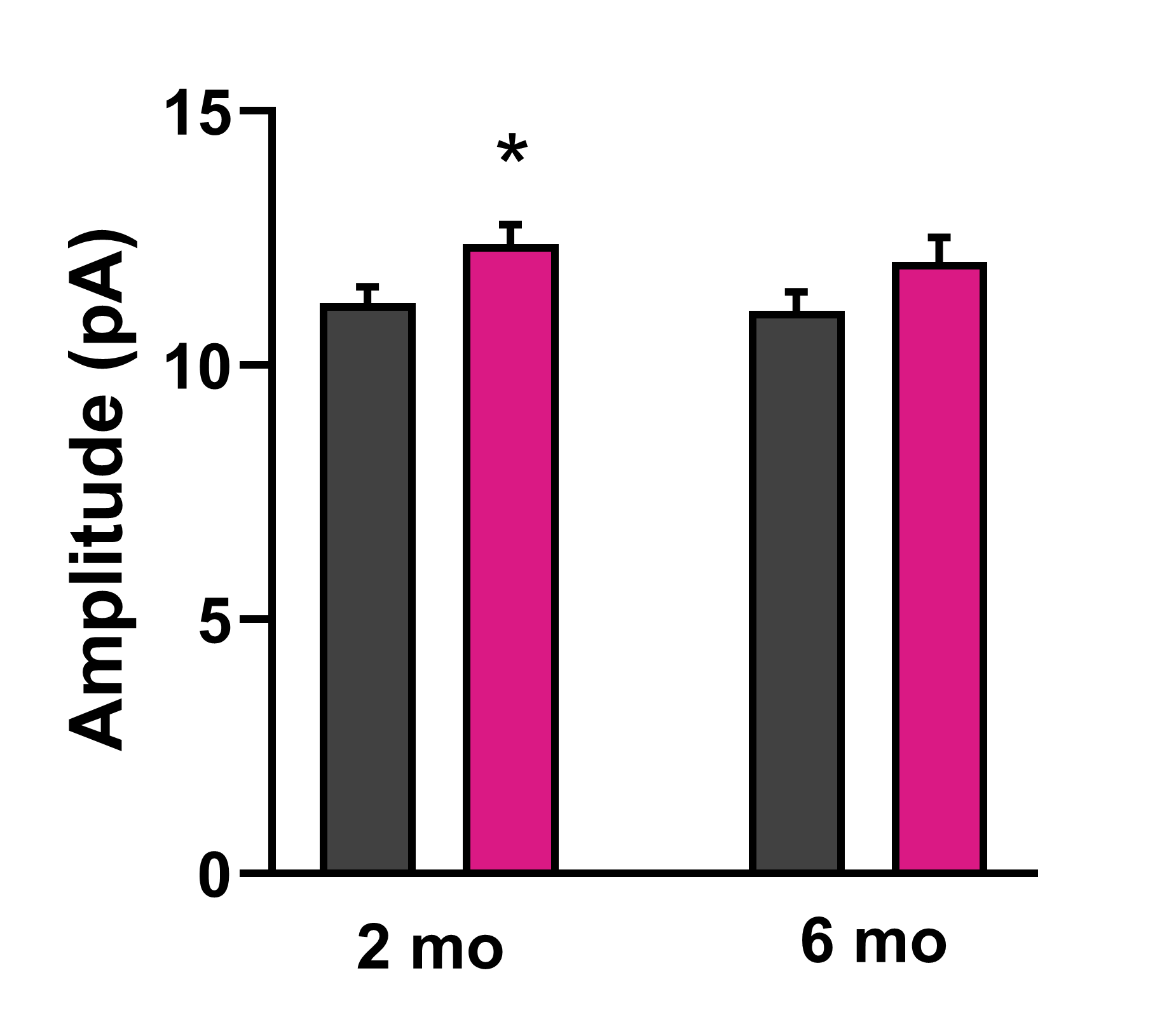
Reductions in mEPSC frequency at corticostriatal synapses observed in Line61 mouse model of Parkinson’s disease
Services
Services
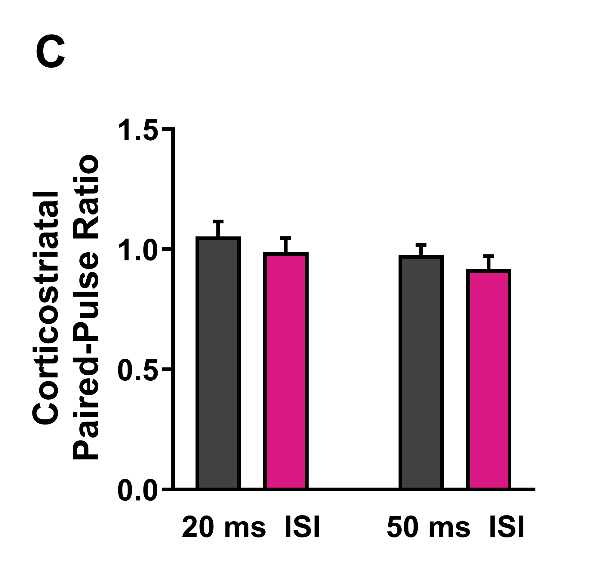
Analysis of glutamatergic synaptic transmission of striatal SPNs in the dorsal striatum using whole-cell patch clamp electrophysiology in brain slices. Brain slices were prepared from Line 61 male mice at 2 and 6 months of age and mEPSCs were recorded (in the presence of 1 μM tetrodotoxin and 40 μM picrotoxin) (n=30-48 neurons from 6 mice per genotype). (A) Frequency of mEPSCs was significantly reduced in Line 61 mice as early as 2 months of age, ****p<0.0001. (B) Amplitude of mEPSCs was slightly increased in Line 61 mice as a potential compensatory mechanism for reduced mEPSC frequency (*p<0.05). (C) Corticostriatal release probability assessed by paired pulse stimulation of cortical afferents (20 and 50 ms inter-stimulus intervals; ISI) was unaltered.