

Services
Services
Electromyography (EMG) is a diagnostic technique which utilizes surface or subdermal electrodes placed at specific sites to optimally stimulate motor and/or sensory nerves to produce muscular contractile /nerve firing responses. Analysis of these responses allows for assessment of nerve (CAP – compound action potential) and muscle (CMAP – compound muscle action potential) function and quantitative assessments of neuromuscular pathology. EMG offers a minimally invasive, repeatable approach that can be used to evaluate longitudinal changes in nerve and muscle function. Psychogenics’ deployment of this technique has been used to produce reliable, consistent, and translatable results in preclinical animal models of neurodegenerative/ neuromuscular diseases and peripheral neuropathies, including (but not limited to) ALS and Charcot-Marie Tooth disorders.
ALS mouse models
TDP-43 ΔNLS Mice Exhibit Inducible Progressive Neuromuscular Deficits
Services
Services
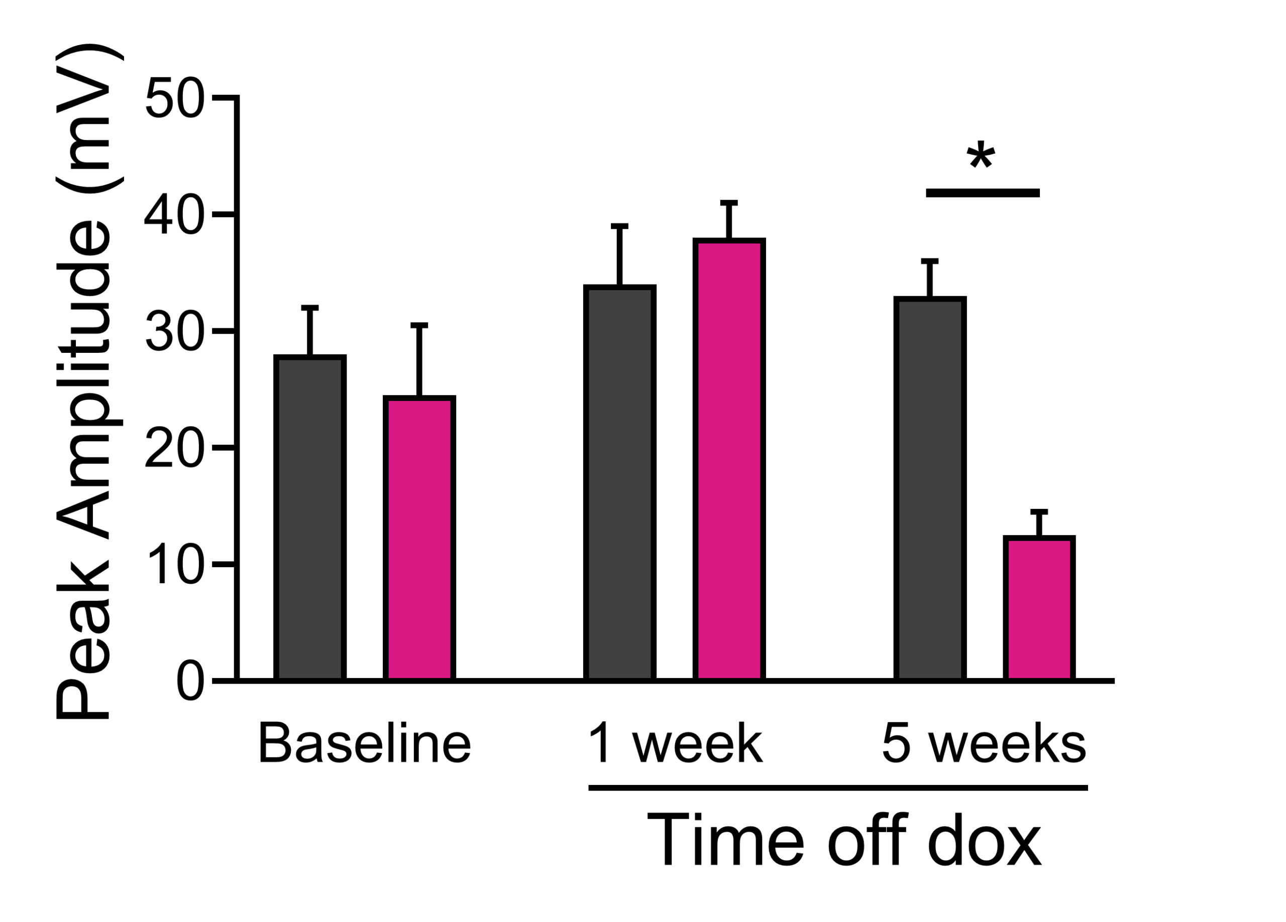
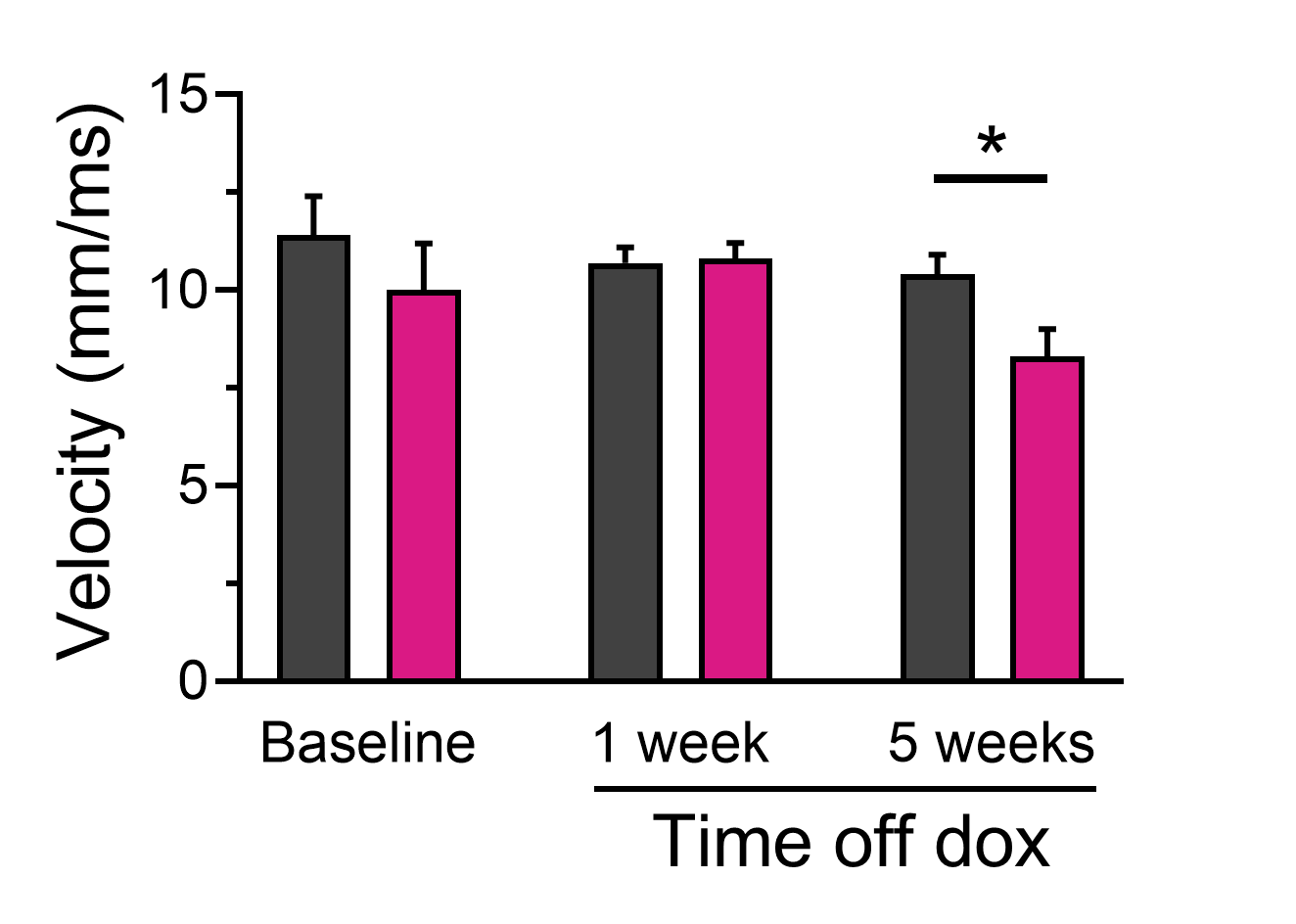
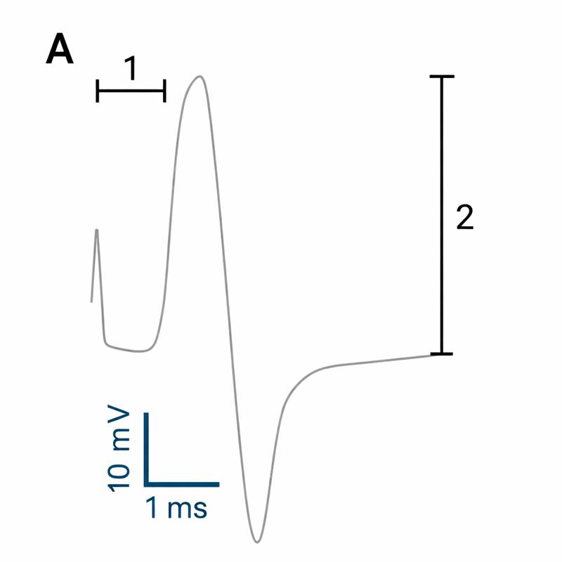
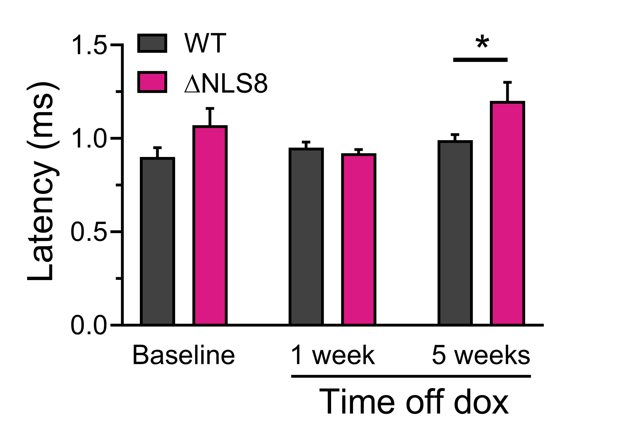
The TDP-43 DNLS mouse model of ALS is doxycycline-regulatable; maintaining animals on doxycycline (dox) suppresses the development of ALS pathology, which progresses once dox is removed. Here we show CMAP responses in gastrocnemius muscle recorded via EMG in combined male and female WT and ΔNLS mice, while on (Baseline) and following removal of dox-containing chow at 5 weeks of age. Deficits in measured parameters appear by 5 weeks following removal of dox. (A) Illustrative CMAP trace indicating measurement of key variables. 1 = response latency, the delay between stimulus of the afferent motor nerve and target muscle contraction. 2 = peak response amplitude, the ideal maximum of which represents full muscle activation by the motor nerve. (B) response latency, showing increasing delays at 5 weeks off dox. (C) muscle response amplitude, showing major functional loss (consistent with observed muscle wasting in ALS) at 5 weeks off dox. (D) Neuromuscular conduction velocity, which combines both nerve conduction velocity and the delay at the neuromuscular junction.
TDP-43 Q331K Mice Exhibit Slower-Developing Milder Neuromuscular Deficits
Services
Services
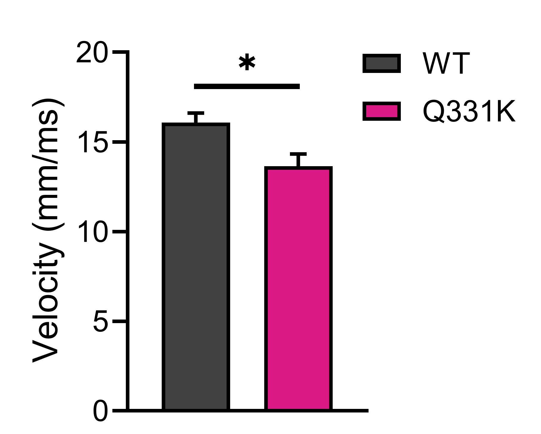
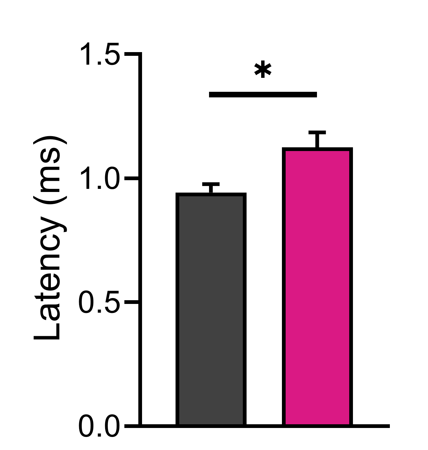
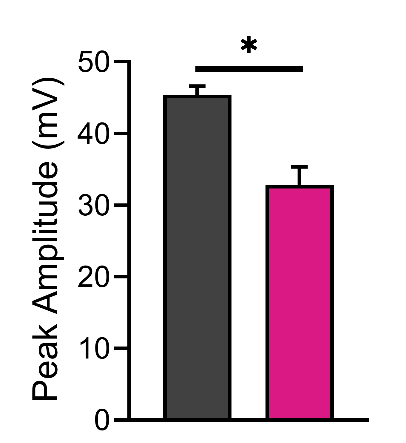
The TDP-43 (Q331K) mouse model of ALS produces age-dependent progressive degeneration of lower motor neurons, producing adult-onset motor deficits. Here we show analysis of CMAP responses in gastrocnemius muscle recorded from mixed male and female WT and TDP-43 Q331K mice at 9 months of age. (A) Response latency was significantly impaired in TDP-43 Q331K mice. (B) Peak response amplitude was significantly reduced in TDP-43 Q331K mice compared to WT. (C) Neuromuscular conduction velocity was slower in TDP-43 Q331K mice.
SOD1 (G93A) mice develop a severe neuromuscular phenotype consistent with advanced muscle atrophy
Services
Services
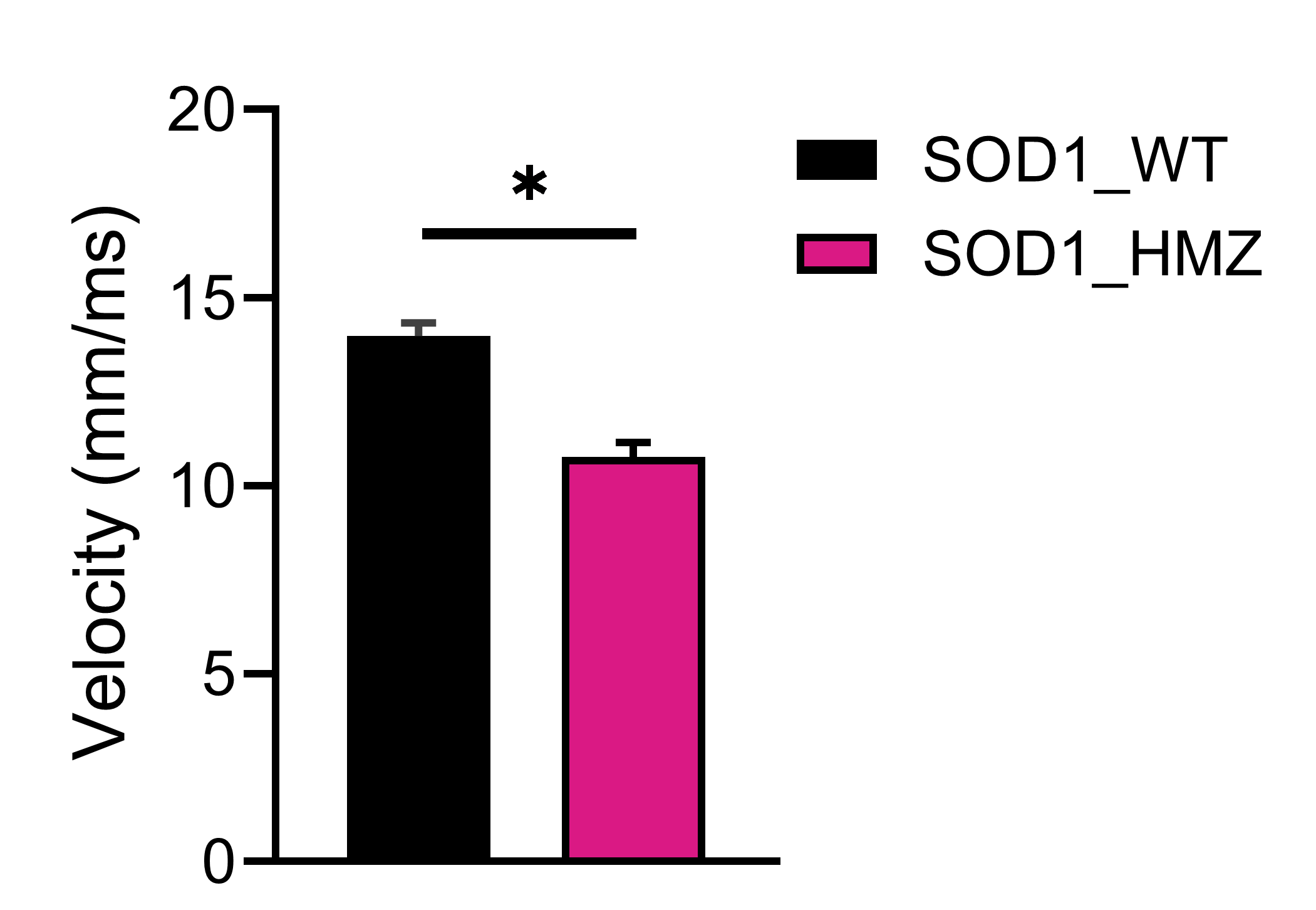
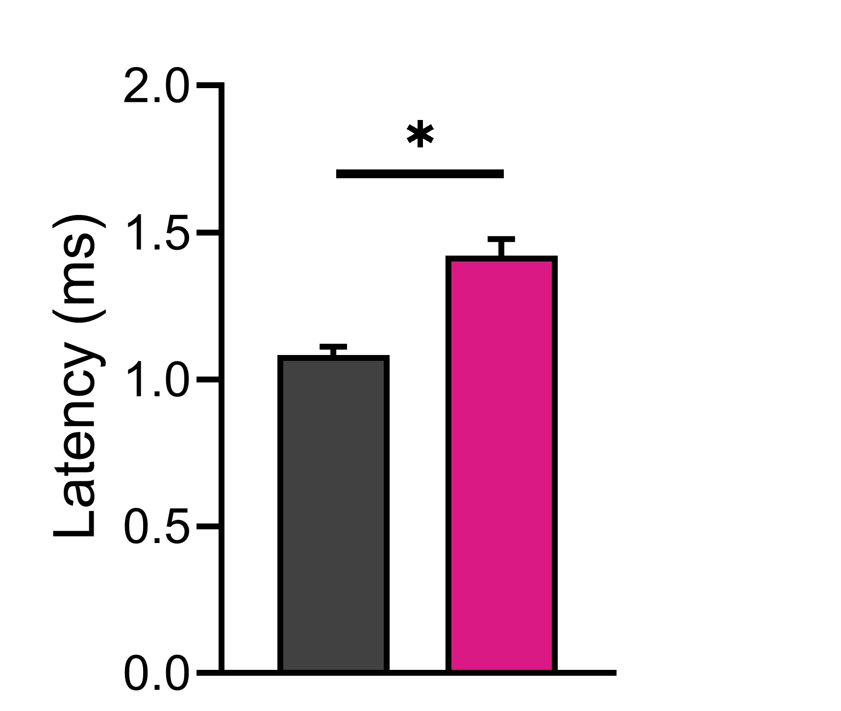
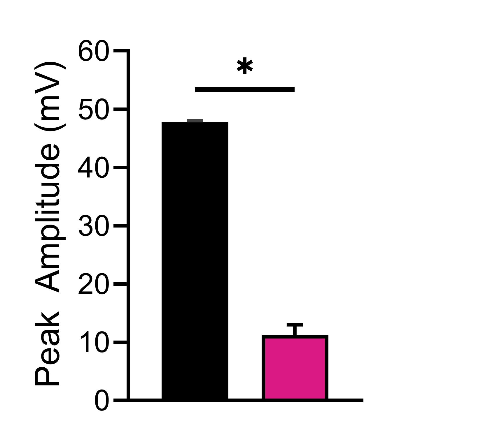
The SOD1 (G93A) transgenic mouse model of ALS expresses the human SOD1 gene, producing a phenotype similar to the symptomology observed in human ALS patients. Here we show analysis of CMAP responses in gastrocnemius muscle recorded from female WT (non-carrier) and SOD1 (G93A) mice at 18 weeks of age. (A) Response latency was significantly impaired in SOD1 (G93A) mice. (B) Peak response amplitude was significantly reduced in SOD1 (G93A) mice compared to WT, reflecting a dramatic loss of muscle mass. (C) Neuromuscular conduction velocity was slower in SOD1 (G93A) mice.
Charcot Marie Tooth
C3-PMP22 Mice Have Diminished Neuromuscular Performance
Charcot-Marie-Tooth disorders are peripheral neurodegenerative diseases, of which CMT1A is the most common form; it is caused by duplication of the peripheral myelin protein 22, which results in demyelination of peripheral nerves.
Services
Services
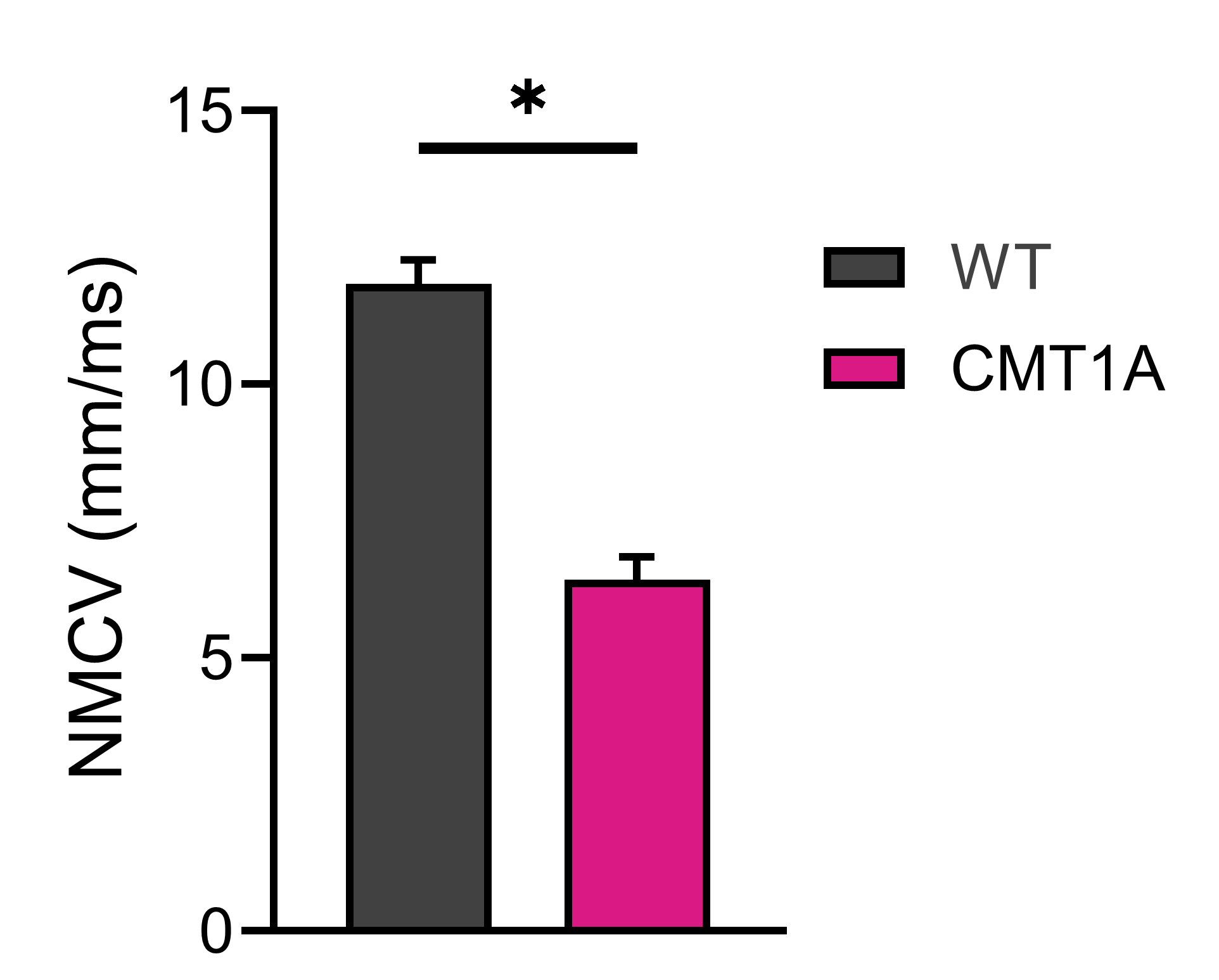
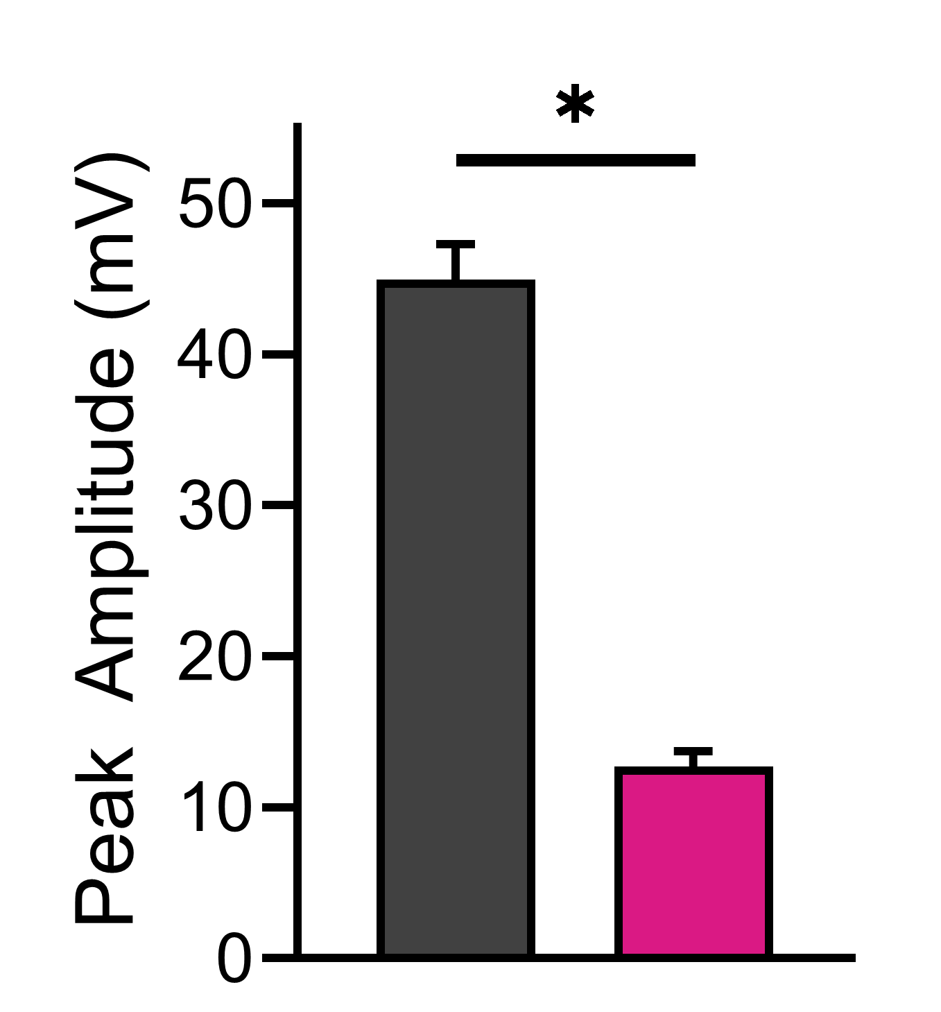
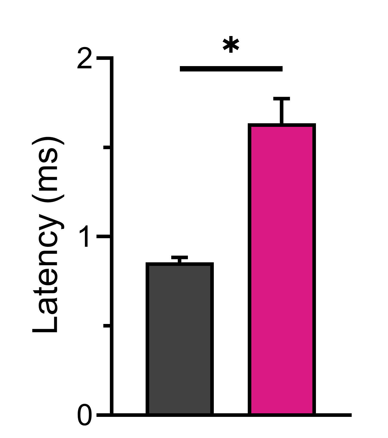
CMAP responses in gastrocnemius muscle recorded from 14-week-old WT and C3 mice show that: (A) Peak response amplitude was significantly decreased in CMT1A mice compared to WT. (B) Response latency was significantly impaired in CMT1A mice. (C) Neuromuscular conduction velocity was severely compromised.
CMT1A Rats Display Impaired Sensory Nerve Conductance
Services
Services
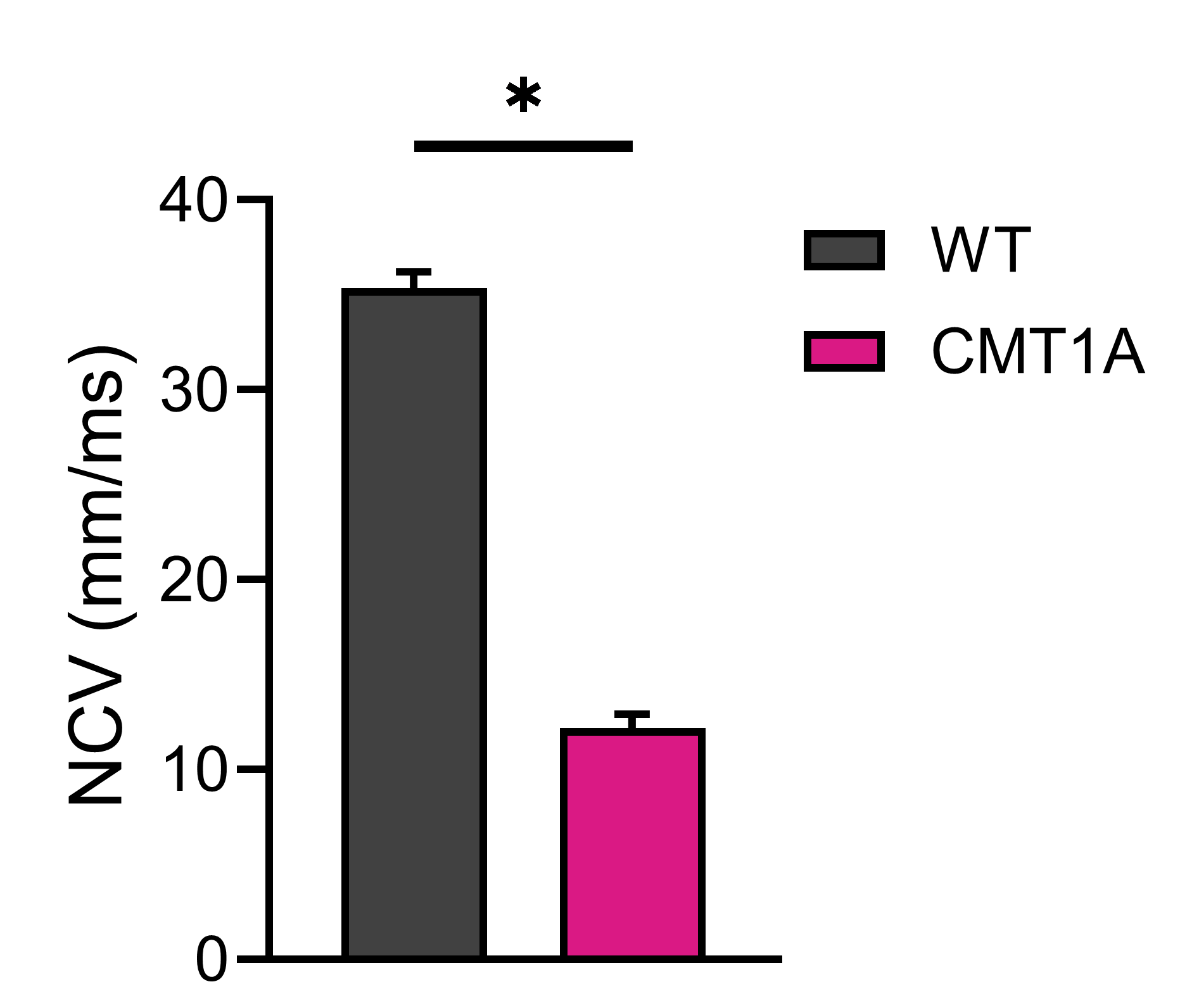
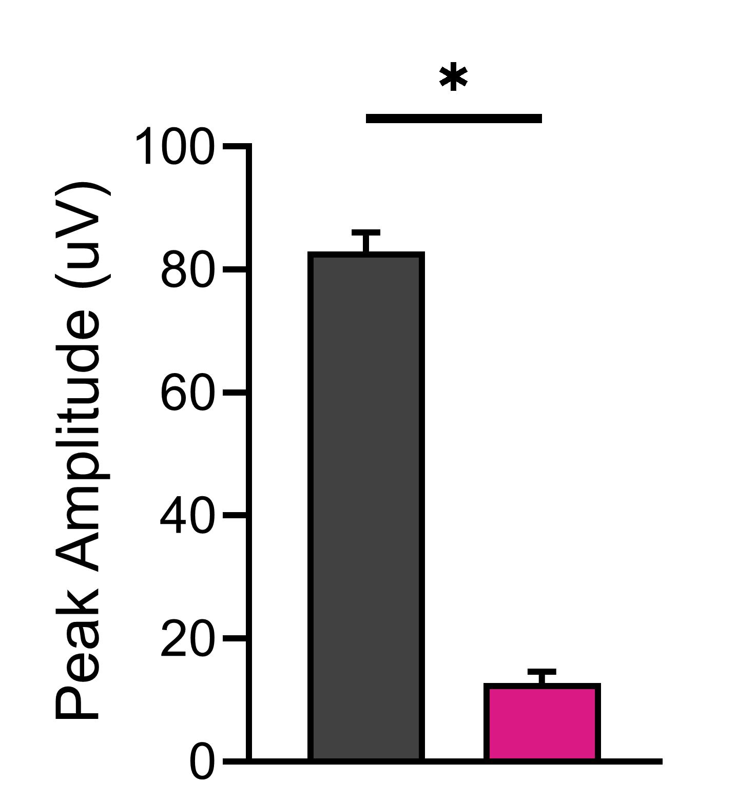
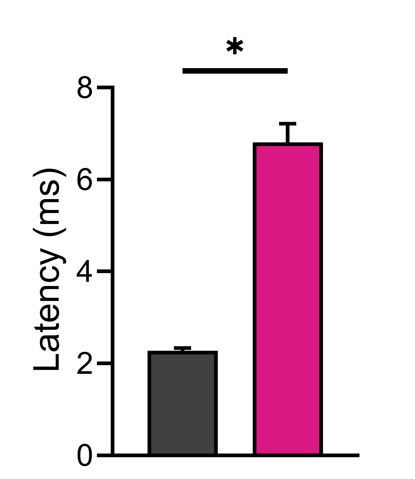
The demyelination of peripheral nerves in CMT1A results in clearly discernable nerve conduction deficits which can be examined directly using CAP analysis of sensory nerve responses in the tails of mice or rats. Here we show CAP analysis of 17-week-old WT and CMT1A male rats. (A) Peak response amplitude was significantly lower in CMT1A rats compared to WT. (B) Response Latency was significantly delayed in CMT1A rats. (C) As may be expected as a consequence of demyelination, nerve conduction velocity was severely compromised in CMT1A rats.
Parkinson’s Disease
Line 61 Mice Exhibit Neuromuscular Fatigue
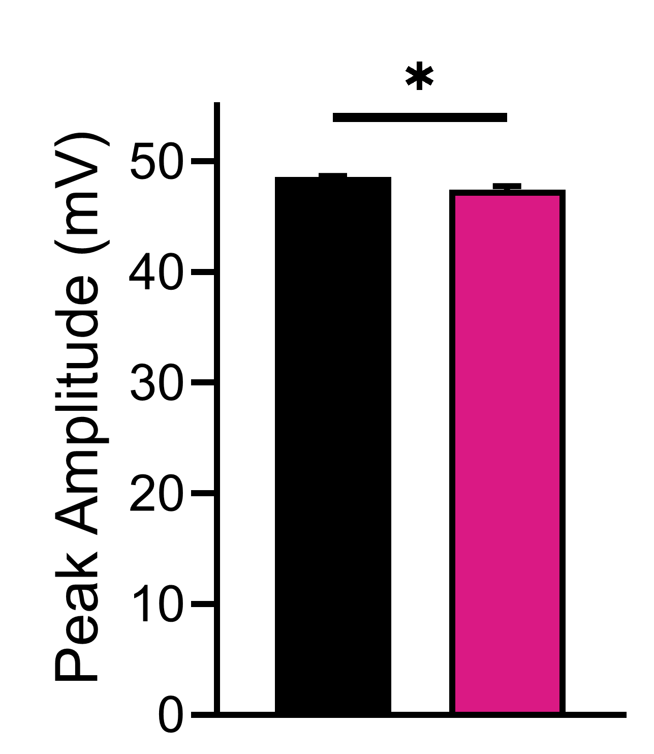
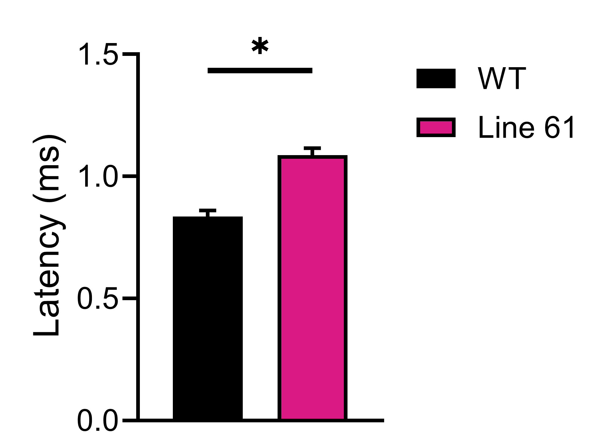
The Line 61 mouse model of Parkinson’s disease exhibits numerous aspects of the human disease, including deficits in muscle strength. We measured CMAP responses in gastrocnemius muscle in response to sciatic nerve stimulation in 26-week-old male Line 61 and ~30-week-old male WT (C57/Bl6) mice.
Services
Services
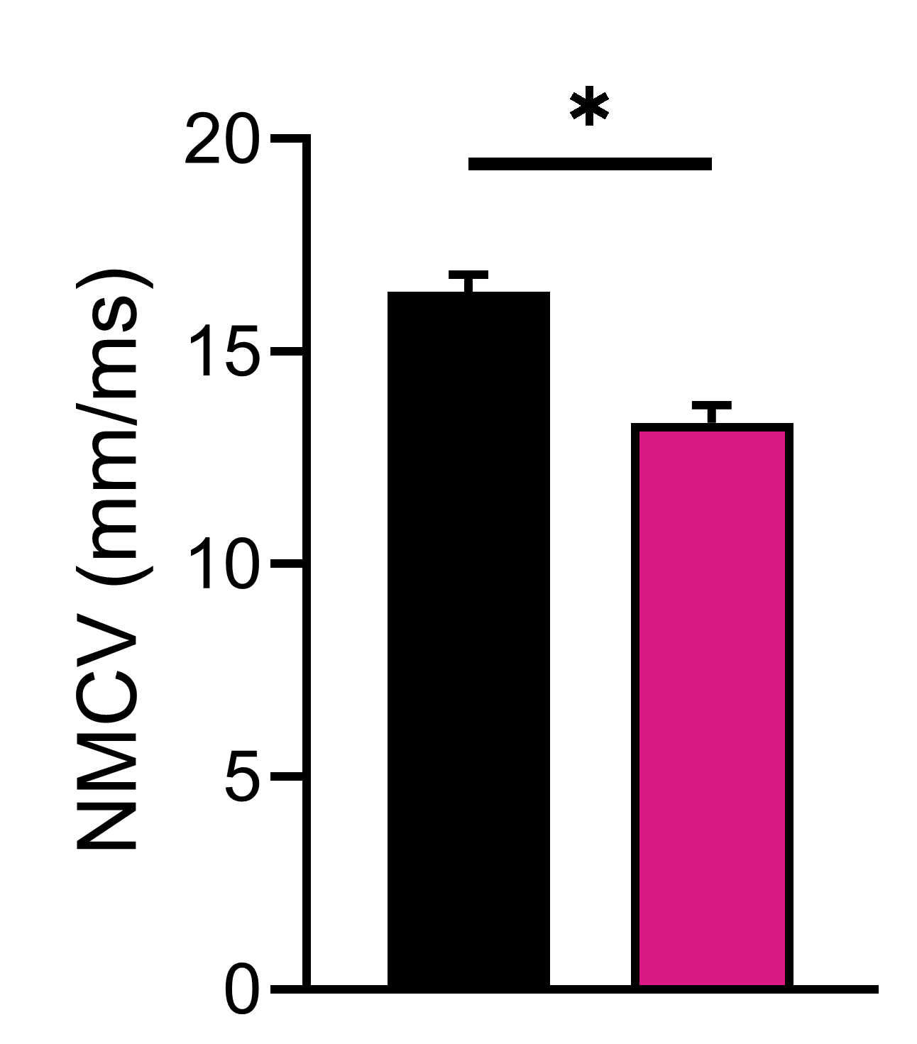
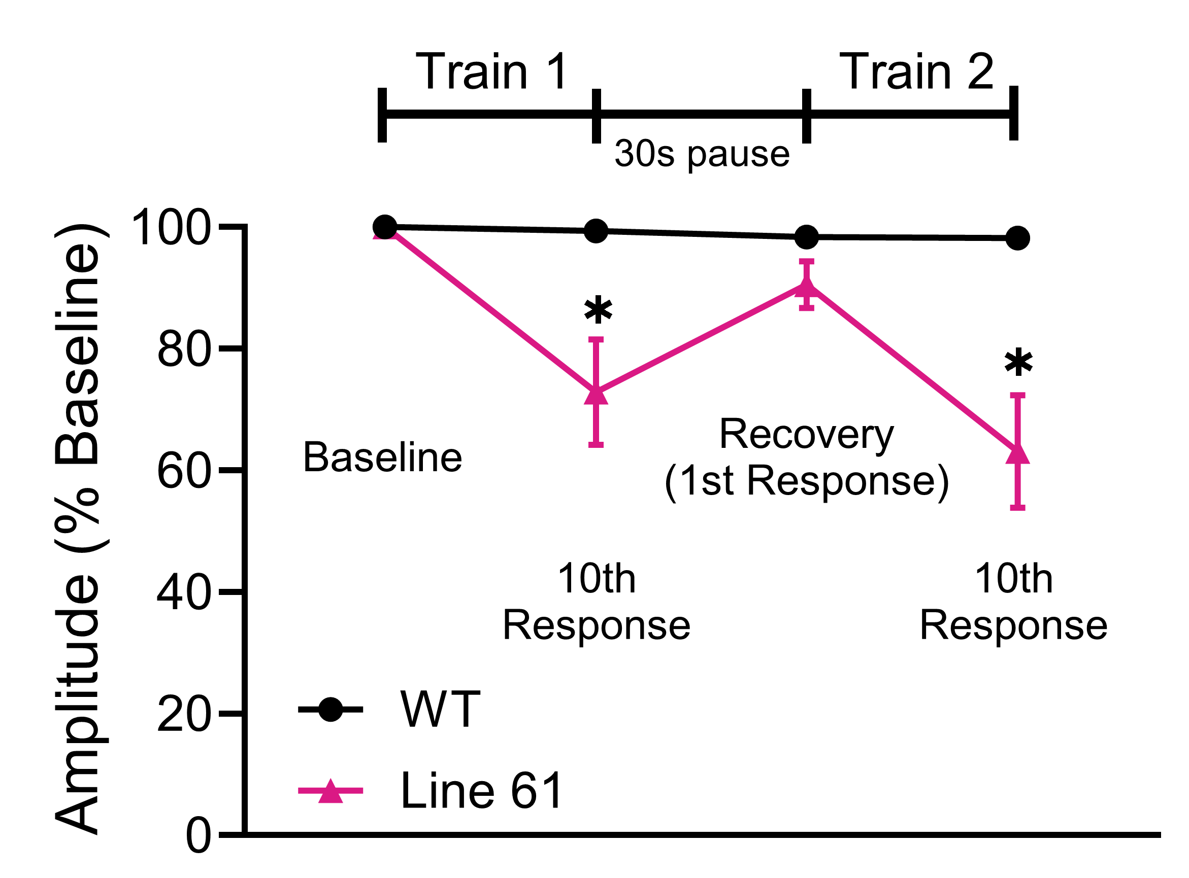
A) Line 61 animals have a slightly decreased muscle response amplitude, although this is likely not biologically relevant. (B) Line 61 animals had a delayed muscular contractile response to motor nerve stimulation. (C) Similarly, neuromuscular conduction velocity (NMCV) was significantly impaired in Line 61 animals. (D) In a muscle fatigue EMG assay, Line 61 animals showed significantly decreased response amplitudes during repetitive stimulation of the sciatic nerve, whereas WT animals showed little to no fatigue in response to the same stimulation.