PsychoGenics leverages transgenic models to advance preclinical studies of amyotrophic lateral sclerosis (ALS), a progressive paralytic disorder characterized by motor neuron degeneration in the brain and spinal cord. Our expertise extends to both sporadic (SALS) and familial (FALS) forms of ALS, with a particular focus on mutations in the superoxide dismutase 1 (SOD1) and TDP-43.

The SOD1 (G93A) mouse model is used to study Amyotrophic Lateral Sclerosis (ALS), a neurodegenerative disease affecting motor neurons. These mice overexpress a mutant human SOD1 gene, leading to a phenotype that mimics human ALS symptoms, including changes in body weight, declining grip strength, and reduced motor activity. This model has been crucial in advancing our understanding of ALS pathogenesis and in testing potential treatments.

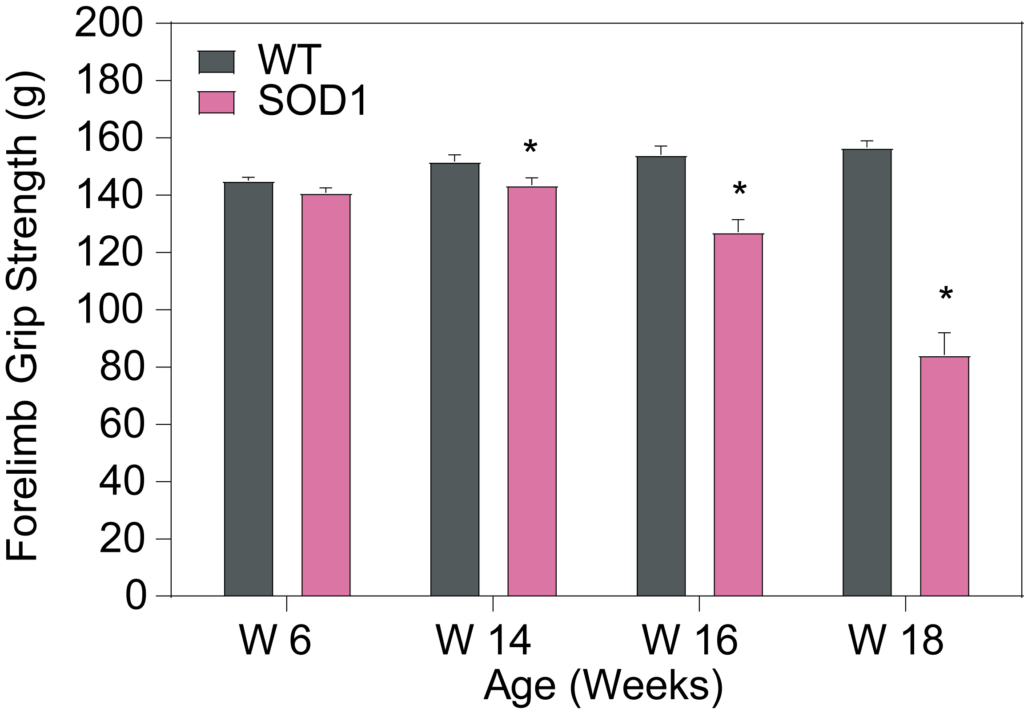
(Left) Progressive decrease in body weight with disease progression. (Middle) Forelimb and (Right) hindlimb grip strength is significantly weaker in SOD1 mice when compared to their WT counterparts.

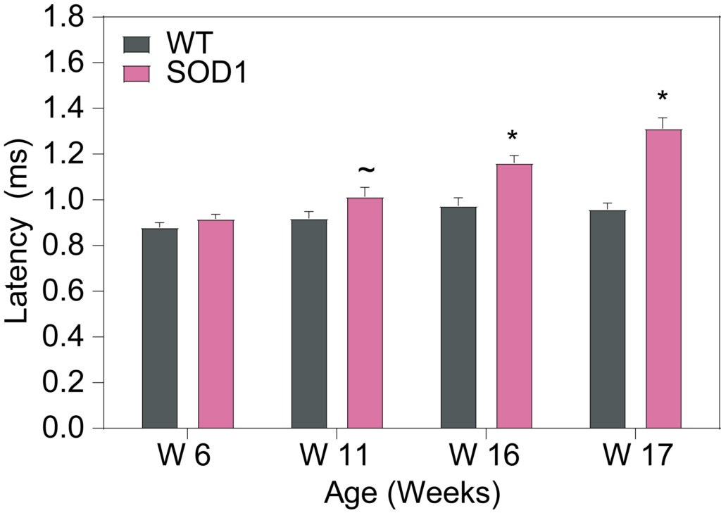
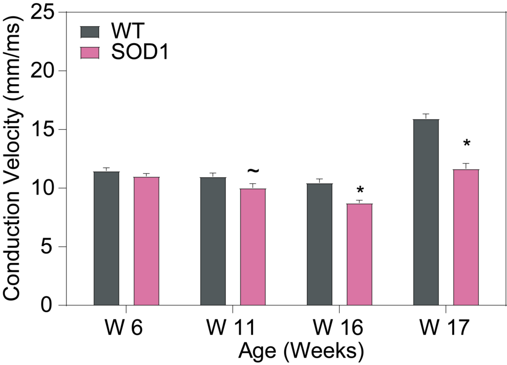
Compound Muscle Action Potential (CMAP) recordings in gastrocnemius. (Left) Response latency is increased, (Middle) CMAP response amplitude is decreased, and (Right) Neuromuscular conduction velocity is decreased, in SOD 1 mice compare to WT controls with disease progression.

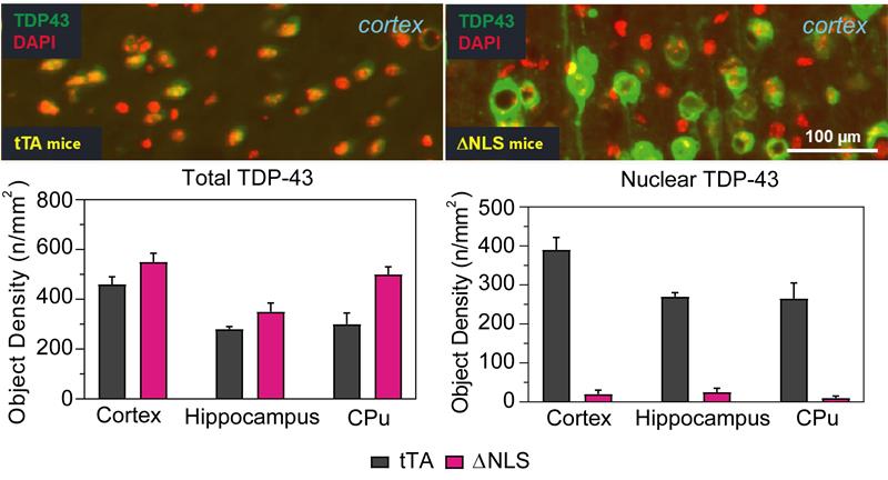
TDP-43 Sub-cellular Distribution – Total TDP-43 is greater in NLS mice compared to tTA mice. Nuclear TDP-43 signals are decreased in NLS mice, as TDP-43 has localized to the cytoplasm.

We characterized the ΔNLS8 inducible TDP43 mouse model of ALS. Upon withdrawal of doxycycline, these mice exhibit progressive motor impairment, muscle denervation, and motor neuron loss, closely resembling the disease pathology observed in ALS patients.

Multiple endpoints have been characterized in the model, including body weight, onset of tremor, clasping behavior, survival, compound muscle action potential (CMAP), gait analysis, and brain and CSF biomarkers (including inflammatory markers and NF-L).

Body Weight and  Survival

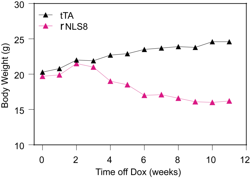
Progressive weight loss in the ∆rNLS8 mice once Dox is removed.

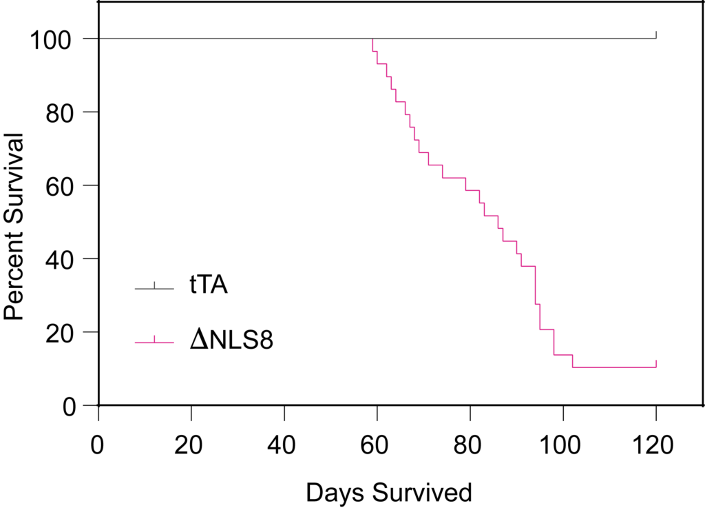
Survival is decreased in the ∆rNLS8 mice after DOX removal.

Tremor and Hindlimb Clasping

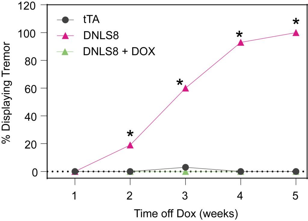
rNLS8 mice show tremors following DOX removal. These tremors are absence when mice are kept on DOX chow.

Motor Deficits

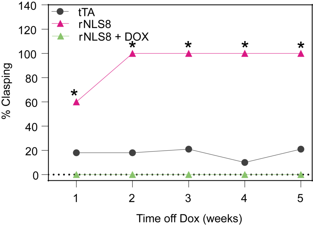
rNLS8 mice exhibit hindlimb clasping after DOX removal. This behavior is not observed in the presence of DOX.

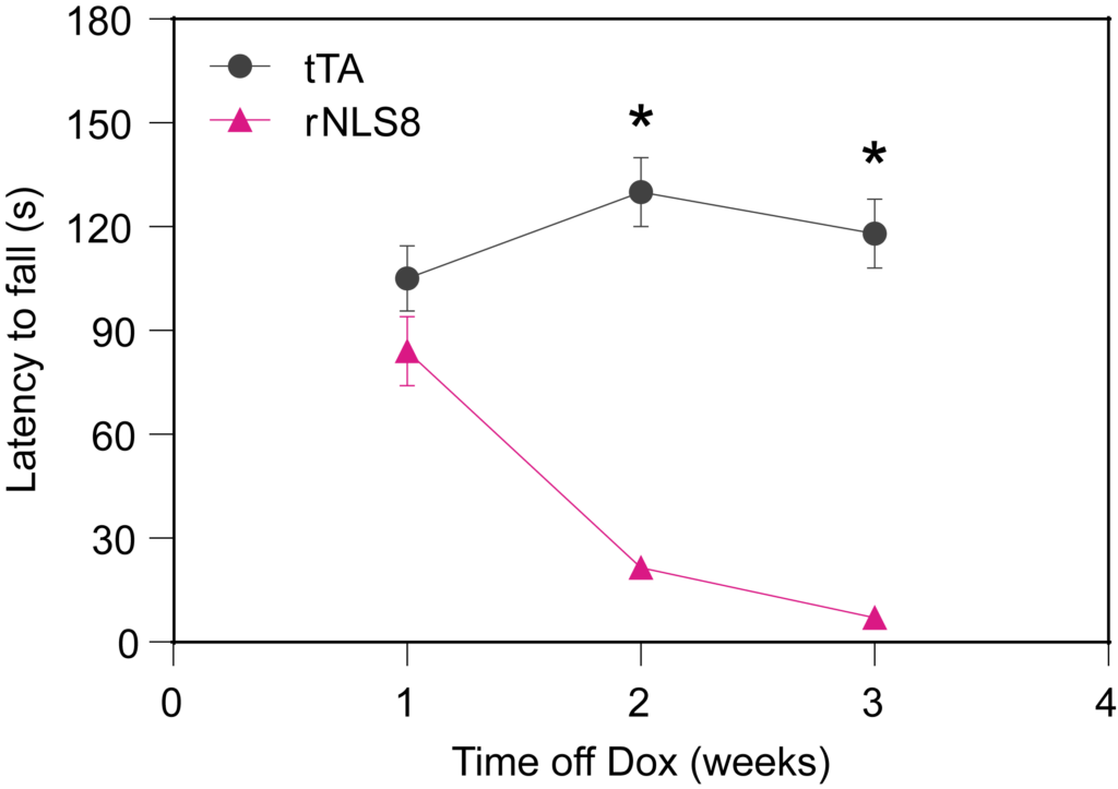
rNLS8 mice show progressive weakness in the wire hang test and fall quicker compared to the tTA mice. Deficits are seen within 2 weeks after DOX removal.

NLS8 mice fall quicker from a rotating rod compared to tTA mice. The deficits in rotarod performance is seen as early as 1 week post DOX removal.

CMAP

rNLS mice show an increase in the onset latency of muscle activation in response to motor nerve stimulation4 weeks post DOX removal.

rNLS mice show a decrease in response amplitude 4 weeks post DOX removal.

rNLS mice show a decrease in muscle nerve conduction 4 weeks post DOX removal.

Neurofilament Light Chain (NF-L)

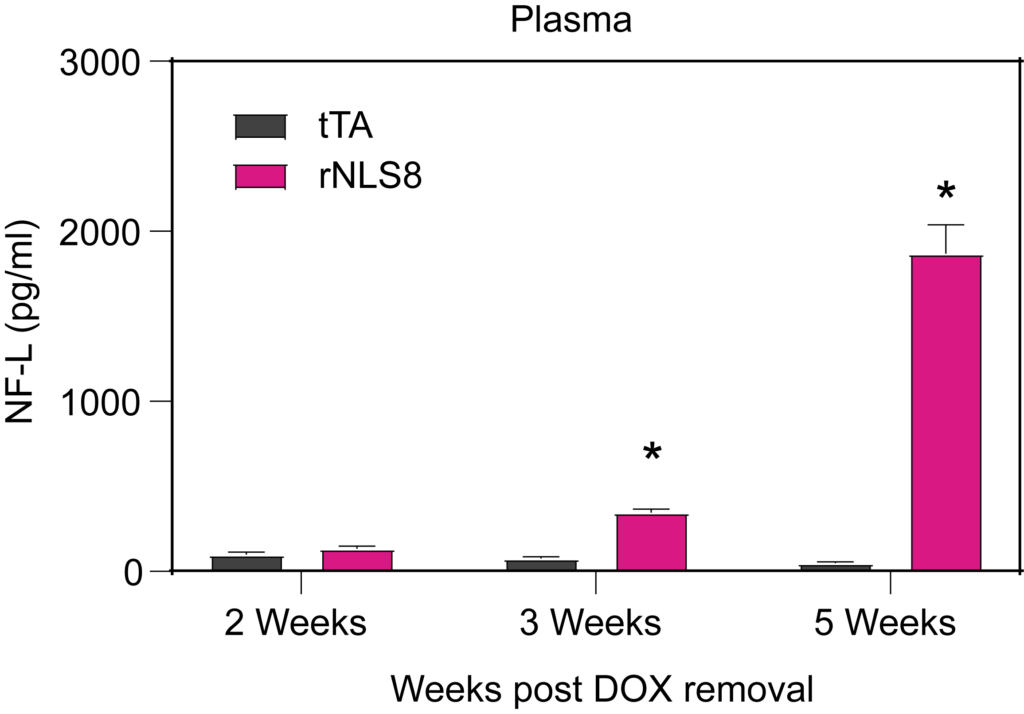
rNLS8 mice show age-dependent elevation of plasma NF-L levels following cessation of DOX treatment.

rNLS8 mice show age-dependent elevated CSF NF-L levels following cessation of DOX treatment.

Subcellular Distribution

TDP-43 Sub-cellular Distribution – Total TDP-43 is greater in NLS mice compared to tTA mice. Nuclear TDP-43 signals are decreased in NLS mice, as TDP-43 has localized to the cytoplasm.

Partnering for Preclinical ALS Research Breakthroughs

PsychoGenics actively collaborates with researchers, pharmaceutical companies, and foundations committed to ALS research. Our extensive experience and specialized models provide a comprehensive and tailored approach to the preclinical development of your novel ALS therapy. 

Collaborate with us to deepen our understanding of ALS pathogenesis and advance your therapeutic breakthrough, ultimately enhancing the lives of ALS patients worldwide.

Partner with PsychoGenics

and discover your next breakthrough

Get Started