The APP/PS1 mouse model expresses mutated forms of the human amyloid precursor protein (APPsw) and presenilin 1 (m146L), facilitating the study of progressive amyloid deposition and neurodegeneration and offering valuable insights into disease progression.
Behavioral Assessments in APP/PS1 Mice
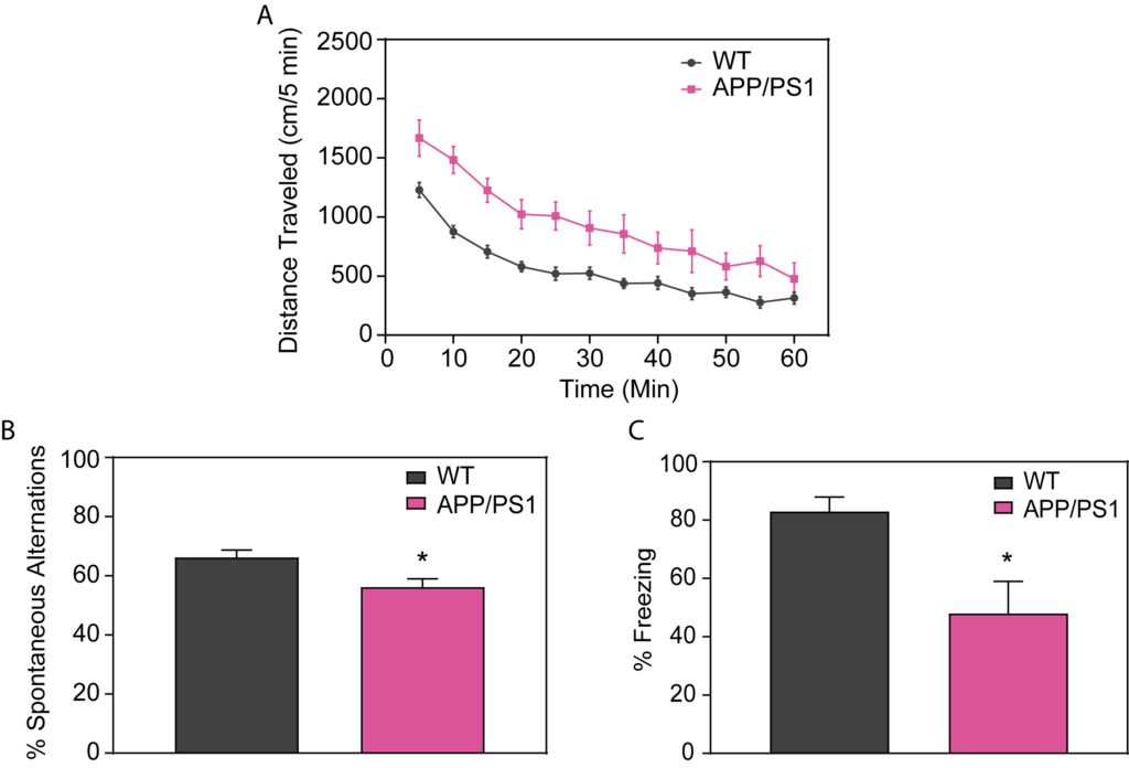
Female APP/PS1 mice show hyperactivity in the open field test (A). These mice exhibit cognitive deficits as seen in the decreased percent alternation in the Y-maze (B) and in contextual fear conditioning as measured by their decreased freezing response (C) compared to WT mice.
APP/PS1 Mice Exhibit LTP Deficit
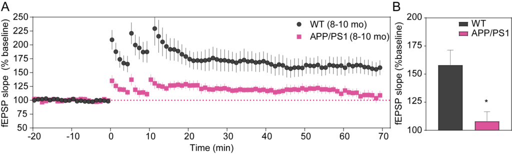
A) A time-course of evoked responses (fEPSP slope normalized to the baseline) showing hippocampal LTP deficit in in 8-10-month-old APP/PS1 mice. (B) Summary of data taken from the last 5 minutes of recordings. Presynaptic axonal excitability appears normal in APP/PS1 mice at this age, suggesting an impairment in synaptic function underlies the observed reduction in LTP (Gelman et al., 2018; J. Alzheimer’s Dis. 61:195-208).
Neuronal Loss and Gliosis in APP/PS1 Mouse Model
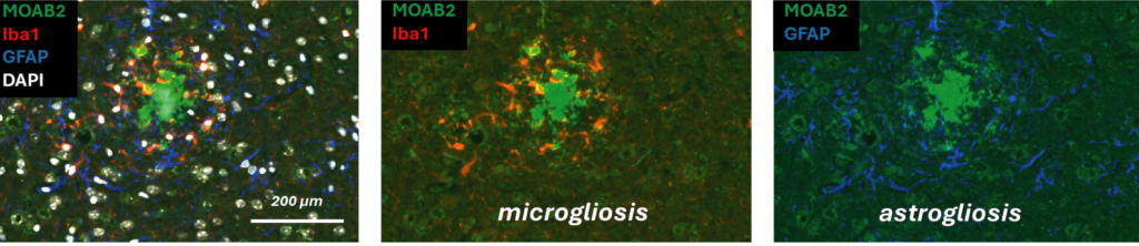
APP/PS1 mice are characterized by a strong and progressive amyloid pathology with concomitant astrogliosis, microgliosis, synapse, and neuronal loss. The images above illustrate a mature neuritic plaque in the cerebral cortex labeled with MOAB2 against Abeta (green), Iba1 staining microglia (red), and GFAP staining astroglia (blue), and DAPI for nuclei (white).
Cortical Markers of Disease Progression in APP/PS1 Mice
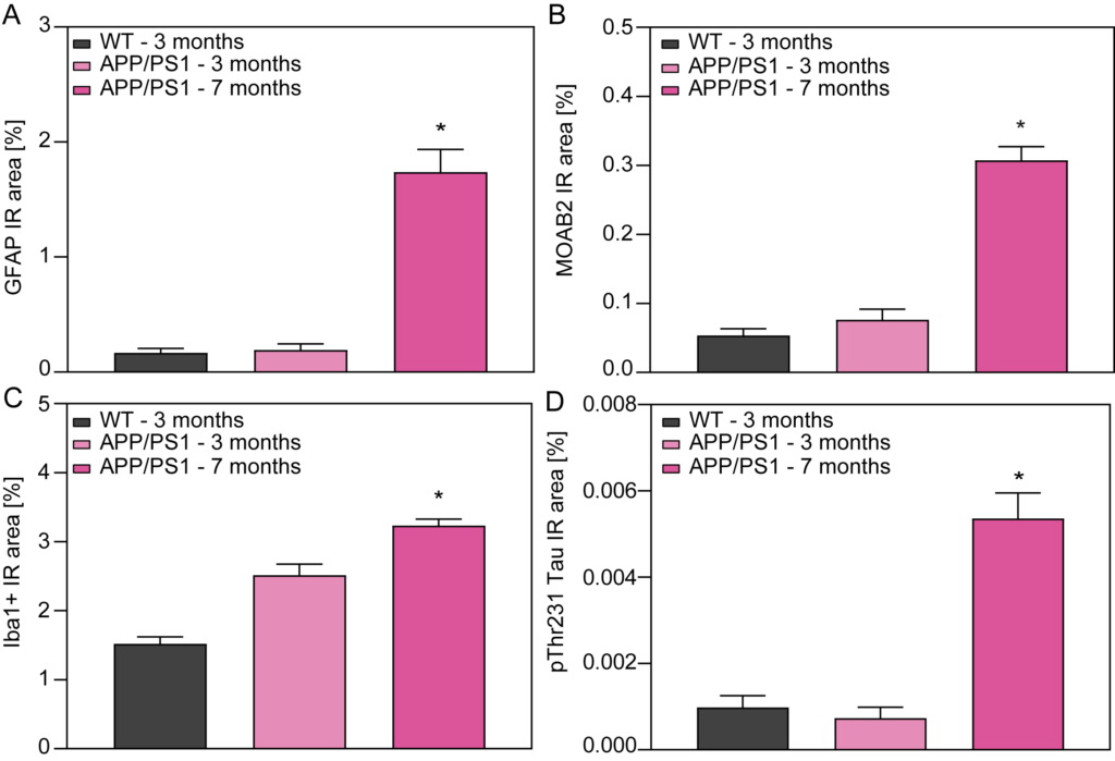
Astrogliosis (A), plaque load (B), microgliosis (C), and phospho tau (D) in cortex of 3- and 7-month-old WT and APP/PS1 mice. Notably, microglial activation can be measured as early as 3 months of age, preceding significant Abeta plaque load. In addition, hyperphosphorylation of Tau at residue Thr231 (and others) is increased in 7-month-old APP/PS1 mice.